|
| Vertices of polygonal epithelial cells: molecular organization of tricellular
tight junctions and their contribution to the epitheliual barrier function |
Epithelial cells in the epithelial cellular sheet are often represented as polygons. In a macroscopic view, tight junctions (TJs) circumscribe each cell as a belt, such that the intercellular spaces within the cellular sheet are continuously sealed. However, TJs cannot practically seal some exceptional regions, namely tricellular contacts, where the corners of three polygonal epithelial cells meet. There are three plasma membranes at tricellular contacts, while TJs are zipper-like structures formed between two plasma membranes. How is the extracellular space at tricellular contacts plugged?
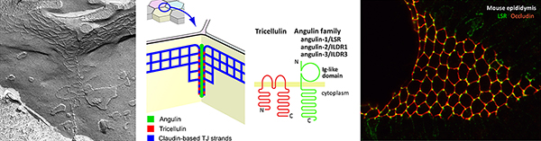
Tricellular contacts have specialized structures of TJs, termed tricellular
tight junctions (tTJs), which extend from apical to basal direction at
the center of tricellular contacts to form very narrow tubes as diffusion
barriers. We identified angulin family proteins, including angulin-1/LSR,
angulin-2/ILDR1 and angulin-3/ILDR2, as molecular components of tTJs. We
demonstrated that angulins are required for full barrier function of the
epithelial sheet and recruit tricellulin, another tTJ-associated membrane
protein, to tTJs. However, detailed mechanism of tTJ formation and functional
difference of angulins in the epithelial barrier function remain elusive.
Moreover, the structure, behavior and physiological function of tricellular
contacts still remain elusive despite their potential significance in epithelial
biology. We address these issues by analyzing the function of the angulin-tricellulin
system by cell biological and physiological approaches.
|
|
|

|
|
| 1. |
Sugawara T, Furuse K, Otani T, Wakayama T, Furuse M. (2021) Angulin-1 seals tricellular contacts independently of tricellulin and claudins. J Cell Biol. 220(9) e202005062. doi: 10.1083/jcb.202005062 |
| 2. |
Oda Y*, Sugawara T*, Fukata Y, Izumi Y, Otani T, Higashi T, Fukata M, Furuse M. (2020) The extracellular domain of angulin-1 and palmitoylation of its cytoplasmic region are required for angulin-1 assembly at tricellular contacts. J Biol Chem. 295:4289-4302. (*equal contribution) |
| 3. |
Higashi T, Katsuno T, Kitajiri S, Furuse M. (2015) Deficiency of Angulin-2/ILDR1, a Tricellular Tight Junction-Associated Membrane Protein, Causes Deafness with Cochlear Hair Cell Degeneration in Mice. PLoS One. 10:e0120674. |
| 4. |
Oda Y, Otani T, Ikenouchi J, Furuse M. (2014) Tricellulin regulates junctional tension of epithelial cells at tricellular contacts via Cdc42. J Cell Sci 127:4201-12 |
| 5. |
Iwamoto N, Higashi T, Furuse M. (2014) Localization of angulin-1/LSR and tricellulin at tricellular contacts of brain and retinal endothelial cells in vivo. Cell Struct Funct. 39:1-8. |
| 6. |
Furuse M, Izumi Y, Oda Y, Higashi T, Iwamoto N. (2014) Molecular organization of tricellular tight junctions. Tissue Barriers 2:e28960. Review |
| 7. |
Higashi T, Tokuda S, Kitajiri S, Masuda S, Nakamura H, Oda Y, Furuse M. (2013) Analysis of the angulin family consisting of LSR, ILDR1 and ILDR2: tricellulin recruitment, epithelial barrier function and implication in deafness pathogenesis. J Cell Sci. 126:966-77. |
| 8. |
Masuda S, Oda Y, Sasaki H, Ikenouchi J, Higashi T, Akashi M, Nishi E, Furuse M. (2011) LSR defines cell corners for tricellular tight junction formation in epithelial cells. J. Cell Sci. 124:548-55. |
|