|
|
|
DEPARTMENT OF INFORMATION PHYSIOLOGYDivision of Sensory and Cognitive InformationThe main purpose of this division is to study the neural mechanisms of visual perception. The human visual system is a complicated parallel and distributed system where several neural structures play different roles, but are still able to generate a unified and integrated precept of the outer world. This system also has sophisticated mechanisms that enable reconstruction of three-dimensional structures from two-dimensional retinal images. To understand the neural substrates of these abilities in our visual system, we are recording neuronal activities from the primary visual cortex and extrastriate visual areas. We are analyzing the stimulus selectivity of neurons to determine the representation of various kinds of visual features, such as color, motion, shape and depth. We are also studying the dynamics of visual information processing in the cortex by analyzing the temporal pattern of neural activities. In addition, to explore the ways in which various visual features contribute to visual perception, psychophysical experiments are conducted in this laboratory.
Staff
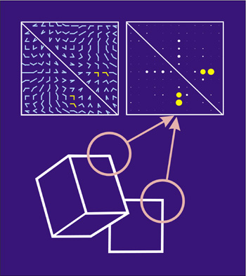
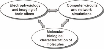
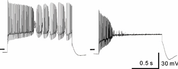
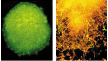
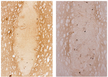
Division of Neural SignalingUsing molecular biological and electrophysiological methods, we investigate the molecular and cellular mechanisms for transduction and integration of information in the nervous system. We are particularly interested in the in vivo roles of neurotransmitter receptors and ion channels. Since molecular genetic analyses have revealed that mutations of functionally critical molecules cause neurological disorders, we also analyze the effects of mutations on the functions of affected molecules, neurons and neuronal circuits. Recently, we have started to make a computational approach, incorporating computer-based neurons into brain slice measurements, together with computational simulation of network functions (Fig.1). The following are currently ongoing major projects. (1) Molecular biological analysis of voltage-gated calcium channels and functional studies of their-associated neurological disorders. Recently, mutations of the voltage-gated calcium channels were found to be associated with neurological disorders of human and mice, which include cerebellar ataxia and some forms of seizure disorders. We try to elucidate the relation how a single mutation can affect brain functions, consequently to result in neurological manifestations, mainly using brain slice preparations (Fig 2). We are also studying the neuronal networks in the cerebellum, the hippocampus and the cerebral cortex. (2) Integration of sensory inputs in the thalamus. The thalamus is located approximately in the center of the brain, and relays the sensory information (pain, temperature, vison, etc) to the cerebral cortex. Recent studies revealed the mechanism of processing the sensory information at the peripheral nerves and the spinal cord, little is known about the operational mechanisms in the thalamus. We have identified PLCb4, which is abundantly expressed in the thalamus, as a key molecule for inflammatory pains. Currently we analyze a model mice lacking the PLCb4 gene, and try to understand the role of thalamic neurons in the sensory processing system. (3) Interaction between heterologous synapses. The synapses, which mediate information between neurons, can be classified into two types; excitatory and inhibitory. Although, in principle, the direction of information in the synapse is uni-directional, recent studies showed that synaptic transmission can be retrograde or heterosynaptic. We found that the excitatory neurotransmitters released from climbing fiber terminals (projecting to cerebellar Purkinje cells from the inferior olive in the brain stem) suppress the inhibitory synaptic transmission from basket cells to Purkinje cells (dis-inhibition). Combination of excitatory input and suppression of inhibitory input seems a refined mechanism to enhance the cerebellar output. We are trying to elucidate the molecular mechanism and physiological significance of the interesting phenomenon.
Staff
Division of Neurobiology and Behavioral GeneticsThe research efforts of this division are focused on determining how mammalian behavior and brain formation are regulated by genetic information. We have developed a gene targeting technique to produce mutant mice using murine embryonic stem (ES) cells. This technique allows us to knock out several genes which are thought to regulate long-term potentiation of synaptic transmission (LTP) and/or neural network formation. The biochemical, histological and behavioral abnormalities in the mutant mice are being examined. Recent interest has focused on mutant mice lacking Fyn, one of the tyrosine kinases in the brain; these animals show impaired in their suckling behavior and spatial learning in the milk pool. To understand the mechanism operating between the absence of Fyn and these behavioral abnormalities, we are seeking to identify the brain regions where the behavior is controlled and the molecules which regulate the behavior in conjunction with Fyn.
Staff
Division of Learning and Memory ResearchIn advanced countries the span of human life has been extended in these decades and the relative population of aged people has been increased. In such societies many people are suffering from various brain dysfunction such as ischemia, Parkinson disease, dementia, Alzheimer disease, and so on. The main research project of this division is "regeneration of nervous system and reconstruction of disturbed brain function". The current aim of research is to study the mechanisms of differentiation of neural stem cells and their development after transplantation in the brain. p037-038(10Neural stem cells proliferate infinitely in serum-free medium supplemented with EGF/FGF and they maintain self-renewing ability over years. Thus, by culturing stem cells, we can get a good amount of cells that would be offered for donor cells in neural transplantation. However, once the neural stem cells were cultured with an ordinary medium with serum, most of them differentiate to astrocytes and only a few to neurons. We have reported that once the mesencephalic stem cells (progenitors) were transplanted in the striata of hemiparkinson model rat, they develop to dopaminergic (DAergic) neurons more extensively in DA-depleted striatum than in intact striatum. This suggests that both genetic background (memory) and environmental cues (conditioning or learning) work together and the final fate of differentiation would be completed. The characterization of the environmental cues that are involved in differentiation of stem cells is now underway using PCR-Select cDNA subtraction, in situ hybridization, SAGE and so on. Another research project of this division is "Functional characterization of Ca2+-permeable channel". Recent molecular biological analyses have revealed the existence of many cation channels in neurons. However, permeation properties, activation mechanisms, and physiological roles of some channels remain unknown. To address these questions, we characterize electrophysiological properties of cloned cation channels and native cation channels in brain neurons.
Staff
|


|
|
|
| Copyright(C) 2004 NIPS (www-admin@nips.ac.jp) | |