|
|
|
CENTER FOR BRAIN EXPERIMENTSection of Brain Structure
|
Staff |
|
 |
Associate Professor: ARII, Tatsuo, PhD 1967 Graduated from Tohoku University, Faculty of Science. Completed the doctoral course in Engineering, Nagoya University. 1972 Research Associate, Nagoya University. 1973 Research Associate, Regensburg University. 1976 Research Associate, Nagoya University. 1979 Associate Professor, NIPS. Speciality: Electron Microscopy |
 |
Assistant Professor: FURUYA, Sonoko, PhD 1970 Graduated from University of Tokyo, Faculty of Pharmacy. Completed the doctoral course in Pharmacy, University of Tokyo. 1975 Research Associate, Nihon Medical College. 1978 Research Associate, NIPS. Speciality: Tissue Culture and Histology |
Section of Brain Function
|
In order to investigate the brain mechanism underlying our mental ability such as cognition, voluntary movement, thinking, or will, we have to experiment on the human brain. Some non-invasive techniques for measuring brain are certainly useful for the purpose. However, they are still insufficient in the quality of information. To overcome the limitations, researches on the brain are carried out here in both the human and monkey subjects using various techniques including direct recordings of cortical field potential, magnetoencephalography, and positron emission tomography.
|
Staff |
|
 |
Associate Professor: TSUJIMOTO, Toru, MD, PhD 1986 Graduated from Kyoto University, Faculty of Medicine. 1990 Completed the doctoral course in Medicine, Kyoto University. 1993 Research Associate, NIPS. 1994 Research Associate, Kyoto University. 1999 Associate Professor, NIPS. Speciality: Neurophysiology |
Section of Information Processing
(Two-photon microscopy)
|
i) Two-photon microscopy imaging group ii) Computer & Network group |
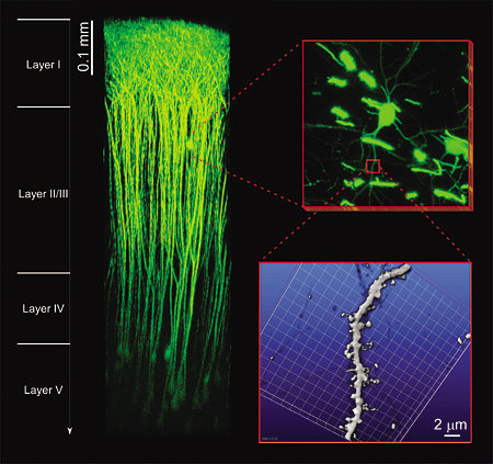 |
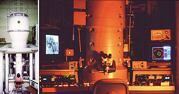
| Figure 1. "In vivo" two-photon microscopy. Superior tissue penetration enables us to monitor neural activity and morphological changes of cells in a brain of a living mouse. Our newly constructed "in vivo" two-photon microscopy can detect fluorescent signals from deeper layers than 0.9 mm from the surface of the brain cortex, so that we can reach neurons in all the layers of the cortex in a living mouse. (Collaborative research with Prof. Junichi Nabekura) |
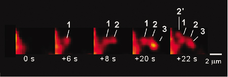 |
| Figure 2. Sequential exocytosis. The deep tissue penetration of two-photon excitation imaging has revealed sequential progression of exocytosis deep into the cytosol in exocrine glands. Now, such sequential compound exocytosis have been found in various kinds of cells, suggesting that it is a general and effective mechanism for physiological secretion (Nature Cell. Biol., 3: 253, 2001). |
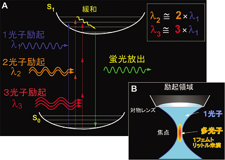 |
| Figure 3. Multi-photon excitation process.Using near infrared femto-second laser, multi-photon excitation of molecules can be elicited by simultaneous absorption of photons (A) at the focal point of an objective lens (B). Two-photon excitation imaging has deep tissue penetration, little out-of-focal light absorption and least phototoxic effects. Thus, it is most suitable for investigating molecular and cellular events within thick intact tissues. In addition, it allows simultaneous multi-color imaging and fluorescence correlation measurement. For example, fusion pore opening and its dynamics can be resolved of a nanometer order by two-photon microscopy (Science, 297:1349, 2002, EMBO J, 25:673, 2006). |
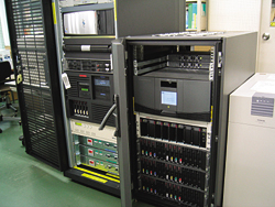 Figure 4. Information processing system |
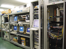 Figure 5. Cluster of network servers |
Staff |
|
 |
Associate Professor: NEMOTO, Tomomi, PhD 1991 Graduated from, Department of Physics, Faculty of Science, the University of Tokyo. 1996 Completed the doctoral course in Applied Physics in Tokyo Institute of Technology. 1996-1997 Frontier Researcher and Special Postdoctoral Researcher, RIKEN. 1997-1999 Research fellow, the University of Tokyo. 1999-2005, Assistant professor, NIPS. 2001-2004, Researcher, PRESTO, JST. Specialty: Cell physiology, Biophysics. |
|
|
|
| Copyright(C) 2006 NIPS (National Institute for Physiological Sciences) | |