DEPARTMENT OF CEREBRAL RESEARCH
The transmission of information in the brain is controlled and regulated by various functional molecules, including receptors, channels and transporters located on the plasma membrane of neuronal and glial cells. The main purpose of this division is to investigate the functional roles of these molecules in the synaptic transmission, neuronal circuits, systematic organization of the brain and animal behaviors, by analyzing their localization, movements, and functions using morphological, electrophysiological, and molecular biological techniques. Special attentions are being made to combine these different techniques efficiently and elucidate the integrated brain functions.
The main projects are as follows.
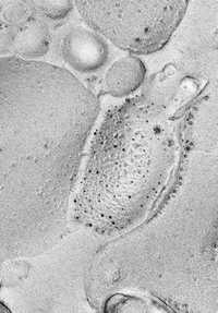
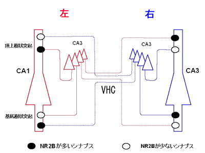
(1) Ultrastructural analysis of the localization of glutamate and GABA receptors, especially in spatial relation to the synapses, and colocalization of these receptors with various channel molecules regulated by receptor activation. Visualization of these functional molecules in the plastic changes, or pathological conditions, using in vitro model systems as well as in vivo. For example, we have recently found colocalization of various subunits of glutamate (Fig.1) and GABA receptors using a newly developed SDS-digested freeze-fracture replica labeling method. This method is highly sensitive and useful for quantification of number and density of receptor and channel molecules. Recently, we are also working on left-right asymmetry of NMDA receptors to clarify its physiological significance and mechanism of its formation.
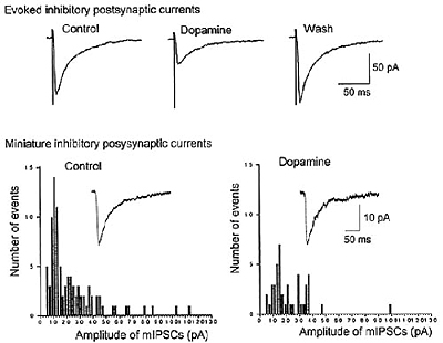
(2) Analysis of synaptic transmission and its modulation in nigro-striatal or mesolimbic dopaminergic system (Fig.2), and cholinergic system in the basal forebrain. These systems are involved in various psychological functions. The regulation of output from these systems to cerebral cortex is also studied.
Another issue is the analysis of pain-perception systems in the spinal cord or brain stem. These studies are carried out using mainly slice-patch-clamp technique. The molecular bases of these mechanisms are also being elucidated.
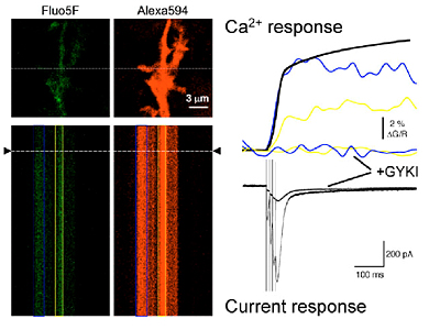
(3) Analysis of the effect of dynamic changes in synaptic and glial microenvironment to the spatiotemporal distribution of neurotransmitters. It has been shown that ectopic release of synaptic vesicles occurs from presynaptic terminals directly facing the surrounding glial cells in the cerebellum (Fig.4). Preliminary data of two-photon imaging show that such form of neural-glial communication may mediate rapid remodeling of the microenvironment. We will analyze how the glial encasement of synaptic structures affects synaptic transmission using combinations of electrophysiological and EM methods.
|
|
|
Fig.1 Co-localization of AMPA-type (5nm gold particles) and GluRd2 (10nm gold particles) glutamate receptors in the cerebellum
|
|
|
|
Fig.2 Asymmetrical allocation of NMDA receptors in the hippocampus
|
|
|
|
Fig.3 Presynaptic inhibition of GABA release in basal ganglia synapses by dopamine
|
|
|
|
Fig.4 Simultaneous recording of Ca increase and current response to synaptic stimuli in glial cells
|
Staff
 |
Professor:
SHIGEMOTO, Ryuichi, MD, PhD
1985 Graduated from Kyoto University Faculty of Medicine. 1986 Resident, Kyoto University Hospital. 1989 Instructor, Kyoto University Faculty of Medicine. 1998 Professor, NIPS.
Speciality: Neuroanatomy and molecular neuroscience |
 |
Associate Professor:
MOMIYAMA, Toshihiko, MD, PhD
1988 Graduated from Kyoto University Faculty of Medicine. 1990 Instructor, Kyoto University Faculty of Medicine. 1994 Postdoctoral Fellow, University College London. 1996 Assistant Professor, Nagasaki University School of Medicine. 1999 Associate Professor, NIPS.
Speciality: Neurophysiology and neuropharmacology |
 |
Assistant Professor:
FUKAZAWA, Yugo, PhD
1988 Graduated from Yokohama City University Faculty of Science. 1997 Completed the doctoral course in Science. 1997 Postdoctoral fellow, Mitsubishi Kagaku Institute of Life Sciences. 2001 Assistant Professor, NIPS.
Speciality: Molecular neuroscience, Endocrinology |
 |
Assistant Professor:
MATSUI, Ko, PhD
1996 Graduated from University of Tokyo, Department of Psychology. 2001 Completed the doctoral course in Psychology at University of Tokyo. 2001 Postdoctoral Fellow at Oregon Health & Science University. 2006 Assistant Professor at NIPS.
Speciality: Neurophysiology |
 |
Postdoctoral Fellow:
SHINOHARA, Yoshiaki, MD, PhD
1996 Graduated from Kyoto University, Faculty of Medicine. 2000 Completed the doctoral course in Medicine at Kyoto University. 2000 Postdoctoral Fellow at Kyoto University. 2001 Postdoctoral Fellow at NIPS.
Speciality: Molecular neuroscience |
 |
Postdoctoral Fellow:
KASUGAI, Yu, PhD
2006 Graduated from School of Life Science, the Graduate University for Advanced Studies. 2006 Postdoctoral Fellow at NIPS.
Speciality: Neuroanatomy |
 |
Postdoctoral Fellow:
TARUSAWA, Etsuko, PhD
2006 Graduated from School of Life Science, the Graduate University for Advanced Studies. 2006 Postdoctoral Fellow at NIPS.
Speciality: Neuroanatomy |
 |
Postdoctoral Fellow:
LÖRINCZ, Andrea, PhD
2000 Graduated from University of Szeged, Faculty of Science. 2004 Completed the doctoral course in Neurobiolgy. 2004 Postdoctoral Fellow at NIPS.
Speciality: Neuroanatomy |
 |
Postdoctoral Fellow:
SÜMEGI, Màté, PhD
2000 Graduated from University of Szeged, Faculty of Science. 2004 Completed the doctoral course in Molecular and Cell Biology. 2004 Postdoctoral Fellow at NIPS.
Speciality: Molecular biolgy |
 |
Postdoctoral Fellow:
KAWAKAMI, Ryosuke, PhD
1997 Graduated from Kyushu University, Faculty of Science. 2003 Completed the doctoral course in Medicine at Kyushu University. 2003 Postdoctoral Fellow at Kyushu University. 2005 Postdoctoral Fellow at NIPS.
Speciality: Neurophysiology |
The neocortex is composed of many functionally-differentiated areas to support the complex activities such as perception, movement and thinking. To understand the function of the cortex, the knowledge of the internal structure of a functional unit in each area is necessary, but not well elucidated yet. Although several types of neurons are involved in the cortical function, the way of information processing in each type of cells and the connection rules among them have not been well understood. Different types of neurons release different chemical substances. How each substance affects the activity of local circuits also need to be understood.
The research in this laboratory concerns the structural and functional analysis of the internal circuits of the cerebral cortex. Physiological characterization of local circuit neurons, functional unit structures in local circuits, and connectional paths among neuronal subtypes will be investigated by electrophysiological, immunohistochemical and morphological techniques to establish the fundamental basis for modeling of the cortical circuitry.
In parallel with functional classification of GABAergic nonpyramidal cells and pyramidal cells projecting to the striatum of basal ganglia, we are investigating the physiological properties of synaptic transmission of each type and their synaptic connections quantitatively in the frontal cortex.
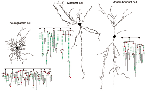
Dendrogram made from dendrites of three cells: LS neurogliaform, non-FS somatostatin Martinotti, and non-FS CRF double bouquet cells. Following are represented with the dendrogram: (1) root issuing primary dendrite (black inverted triangle), (2) node (branching point; red circle), (3) spine (green bar).
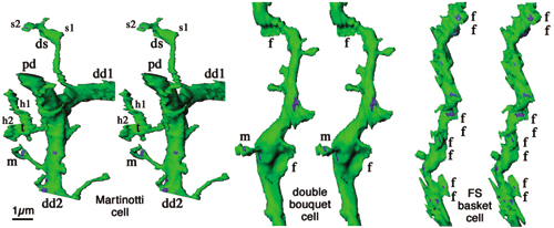
Dendritic structures of Martinotti, double bouquet and FS basket cells. m, mushroom spine; t, triple-branch spine; ds, double-swelling spine; f, fan protrusion. h, spine head, s, spine swelling.
Staff
 |
Professor:
KAWAGUCHI, Yasuo, MD, PhD
1980 Graduated from the University of Tokyo, Faculty of Medicine. 1984 Research Associate, NIPS. 1985 Completed the doctoral course at the University of Tokyo. 1989 Research fellow, RIKEN. 1993 Laboratory head, RIKEN. 1999 Professor, NIPS.
Speciality: Neuroscience |
 |
Associate Professor:
KUBOTA, Yoshiyuki, PhD
Graduated from the master course (1984) and doctor course (1988) at Osaka University, Faculty of Medicine. 1989 Research fellow, University of Tennessee, Dept Anatomy and Neurobiology. 1990 Research Associate, Kagawa Medical School. 1991 Research fellow, RIKEN. 2001 Associate Professor, NIPS.
Speciality: Neuroanatomy, Neuroscience |
 |
Assistant Professor:
OTSUKA, Takeshi, PhD
1997 Graduated from Osaka University, Faculty of Engineering Science. 1999 Graduated from the master course at Osaka University, Graduate School of Engineering Science. 2002 Graduated from the doctoral course at the Osaka University, Graduate School of Engineering. 2002 Research Associate, Duke University Medical Center. 2004 Assistant Professor, NIPS.
Speciality: Neuroscience |
 |
Postdoctoral Fellow:
MORISHIMA, Mieko, PhD
1999 Graduated from Tokyo University of Pharmacy and Life Science.2001 Completed the master course in Osaka University. 2006 Completed the doctoral course in The Graduate University for Advanced Studies. 2006 Postdoctoral fellow, NIPS
Speciality: Neurophysiology |
 |
Postdoctoral Fellow:
GULLEDGE, Allan T, PhD
1991 Received a B.S. in Psychobiology from the University of California, Riverside 2000 Completed a Ph.D. in Biology at the University of Texas, San Antonio. 2000-2005 Research Fellow at the Australian National University
2005 JSPS Postdoctoral Fellowship, NIPS
Speciality: Cellular physiology of neocortical neurons |
The goal of Division of Cerebral Integration is to understand the physiology of human voluntary movement and other mental processing including language using noninvasive functional neuroimaging technique, mainly fMRI. In particular, understanding of the mechanisms of plastic change in the human brain accompanied by learning, sensory deafferentation, and development is the main focus of our research activities. Multimodality approach including EEG, MEG, TMS, and NIR is considered when appropriate.
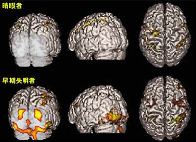
Figure 1.Activation in a sighted (upper row) and blind subject (bottom row) during tactile discrimination tasks similar to reading Braille. The primary and association visual cortices of the blind are activated bilaterally (bottom row) whereas no such activation in the sighted. Only pixels with significant increase in cerebral blood flow during the task were superimposed on surface-rendered high resolution MRI. This is an example of cross-modal plasticity of human brain due to early visual deafferentation and/or long-term training of Braille reading. Depicted by functional MRI using high Tesla (3T) machine.
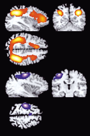
Figure 2 Brain areas for implicit and explicit learning. There are two different way of acquisition of knowledge, implicit learning and explicit learning. Implicit learning is characterized as an unintentional, un-conscious form of learning recognized behavioural improvement (such as shortening of reaction time). Explicit learning involves conscious recollection of previous experiences. In a serial reaction time task, subjects pressed each of four buttons with a different finger of the right hand in response to a visually presented number. Explicit learning was associated with increased activity in the frontoparietal networks (Upper row). During the implicit learning phase, when the subjects were not aware of the sequence, improvement of the reaction time was associated with increased activity in the contralateral primary sensorimotor cortex (Bottom row). These results show that different sets of cortical regions are dynamically involved in implicit and explicit motor sequence learning.
Staff
 |
Professor:
SADATO, Norihiro, MD, PhD
1983 Graduated from Kyoto University School of Medicine. 1994 Completed the doctoral course in Medical Sciences, Kyoto University. 1993-95 Visiting Research Fellow, NINDS, NIH. 1995 Lecturer, Fukui Medical University. 1998 Associate Professor, Fukui Medical University. 1999 Professor, NIPS.
Speciality: Functional neuroimaging |
 |
Assistant Professor:
KANSAKU, Kenji, MD, PhD
1995 Graduated from Chiba University School of Medicine. 2000 Completed the doctoral course in Medical Sciences, Chiba University. 2000-2001 Research Fellow, CREST. 2001-04 Visiting Associate, NINDS, NIH. 2004 Assistant Professor, NIPS.
Speciality: Systems Neuroscience Neuroimaging |
 |
Assistant Professor:
TANABE, Hiroki, PhD
1991 Graduated from College of Liberal Arts, International Christian University. 1998 Completed the doctoral course in Medical Sciences, Osaka University. 1998 Research Fellow, Communications Research Laboratory. 2002 JST Research Fellow. 2004 Assistant Professor, NIPS.
Speciality: Neuroscience and psychology |
 |
Postdoctoral Fellow:
TOYODA, Hiroshi, MD, PhD
1994 Graduated from Faculty of Medicine, Kyoto University. 2002 Completed the doctoral course in Medical Sciences, Kyoto University Graduate School of Medicine. 2003 Postdoctoral Fellow, NIPS.
Specialty: Functional neuroimaging |
 |
Postdoctoral Fellow:
ARAMAKI, Yu, MS
1996 Graduated from Department of Education, University of Tokyo. 1998 Completed the master course, Graduate School of Education, University of Tokyo. 2001 Graduated from the doctoral course, Graduate School of Education, University of Tokyo. 1999 Research Fellow, Research Institute National Rehabilitation Center for the Disabled. 2002 JST Research Fellow.
Speciality: Neuroscience |
 |
Postdoctoral Fellow:
SAITO, Daisuke, PhD
1996 Graduated from Faculty of Integrated Arts and Sciences, Tokushima University. 1998 Completed the master course in Human and Natural Enviroment Sciences,Tokushima University. 2003 Completed the doctoral course in Medical Sciences, Tokushima University. 2002 Fellow, NIPS. 2005, JST Research Fellow.
Speciality: Functional neuroimaging |
 |
Postdoctoral Fellow:
HARADA, Tokiko, PhD
2000 Graduated from College of Humanities and Sciences, Nihon University. 2002 Completed the master course in Integrated Sciences, Nihon University. 2005 Completed the doctoral course in Life Sciences, The Graduate University for Advanced Studies. 2005 Postdoctoral Fellow, NIPS.
Speciality: Functional neuroimaging |
|