DEPARTMENT OF INFORMATION PHYSIOLOGY
The main purpose of this division is to study the neural mechanisms of visual perception. The human visual system is a complicated parallel and distributed system where several neural structures play different roles, but are still able to generate a unified and integrated precept of the outer world. This system also has sophisticated mechanisms that enable reconstruction of three-dimensional structures from two-dimensional retinal images. To understand the neural substrates of these abilities in our visual system, we are recording neuronal activities from the primary visual cortex and extrastriate visual areas. We are analyzing the stimulus selectivity of neurons to determine the representation of various kinds of visual features, such as color, motion, shape and depth. We are also studying the dynamics of visual information processing in the cortex by analyzing the temporal pattern of neural activities. In addition, to explore the ways in which various visual features contribute to visual perception, psychophysical experiments are conducted in this laboratory.
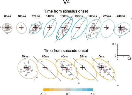
The dominant factor in neural modulations in area V4 dynamically changes from the bottom-up attentional influences to the top-down attentional influences over the course of a visual search. The upper row indicates the neural modulation dynamics after search array presentation and the lower row indicates that before saccade initiation. In the early phase during visual search (upper row), there is positive correlation across data points indicating that bottom-up attention is dominant, whereas in the later phase (lower row), there is negative correlation indicating that top-down attention is dominant. |
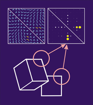
Stimulus selectivity of a monkey V2 neuron to angle stimuli. Top left: angle stimuli were made of a combination of two straight half lines directed toward one of 12 directions. Top right: responses of a V2 neuron to the angle stimulus set. Response amplitude is represented as the diameter of disks. Many V2 neurons exhibited selective responses to particular angles. It is suggested that V2 is the first step to extract information of angles embedded within the contour of objects as shown schematically at the bottom. |
Staff
 |
Professor:
KOMATSU, Hidehiko, PhD
1982 Completed the doctoral course in Osaka University. 1982-1988 Hirosaki University. 1985-1988 National Eye Institute, U.S.A. 1988-1995 Electrotechnical Laboratory. 1995 Professor, NIPS.
Speciality: Neurophysiology |
 |
Associate Professor:
ITO, Minami, PhD
1989 Completed the doctoral course in Osaka University. 1989-1994 Riken Institute. 1994-1998 Rockefeller University. 1998 Associate Professor, NIPS.
Speciality: Neurophysiology |
 |
Assistant Professor:
GODA, Naokazu, PhD
1998 Completed the doctoral course in Kyoto University.1998-2003 ATR. 2003 Research Associate, NIPS.
Speciality: Visual Psychophysics |
 |
Assistant Professor:
OGAWA, Tadashi, PhD
1992 Completed the master course in Osaka University. 1998 received Ph.D. from Osaka University. 1992-1998 Communications Research Laboratory. 1998 Research Associate, NIPS.
Speciality: Control Engineering |
 |
Research Fellow:
KOIDA, Kowa, PhD
2000 Completed the doctoral course in Tokyo Institute of Technology. 2000 Research Fellow, NIPS.
Speciality: Visual Psychophysics |
Our main interest lies in elucidation of the mechanism of transduction and integration of neural information in the nervous system. More specifically, we are trying to understand the basic properties of neural information processing between neurons or among a group of neurons constituting a local network. We are also interested in the pathophysiological mechanism how a single gene mutation leads to a symptom (such as ataxia and epilepsy), particularly in Ca2+ channel mutant mice. Additionally, we have recently started to make a computational approach, incorporating computer-based neurons into brain slice measurements, together with computational simulation of network functions (Fig.1). The following are currently ongoing major projects.
(1) Molecular biological analysis of voltage-gated calcium channels and functional studies of their-associated neurological disorders. Recently, mutations of the voltage-gated calcium channels were found to be associated with neurological disorders of human and mice, which include cerebellar ataxia and some forms of seizure disorders. We study the relation how a single mutation causes neurological manifestations, mainly using brain slice preparations (Fig 2).
Recently, we identified a dramatic impairment in the neural circuit of feedforward inhibition in the thalamocortical projection in epileptic calcium channel mutant mice tottering.
(2) Integration of sensory inputs in the thalamus. Recent studies revealed the mechanism of processing the sensory information at the peripheral nerves and the spinal cord, little is known about the operational mechanisms in the thalamus.
We have identified PLCb4, which is abundantly expressed in the thalamus, as a key molecule for inflammatory pains. Currently we analyze model mice lacking the PLCb4 gene, and try to understand the role of thalamic neurons in the sensory processing system.
(3) Transmitter diffusion-mediated crosstalk between heterologous neurons. In principle, neuronal information is unidirectionally transported at the synapses, however, recent studies suggested that synaptic transmission can be mediated by retrograde and/or heterosynaptic pathways. We reported that the excitatory transmitters diffused from climbing fiber (CF) terminals [projection to cerebellar Purkinje cells (PCs) from the inferior olive in the brain stem] presynaptically suppressed the inhibitory information flow from basket cells to PCs. The heterosynaptic inhibition therefore provides a likely mechanism boosting the CF input-derived excitation to the PCs. We are trying to elucidate the molecular mechanism and physiological significance of this phenomenon.
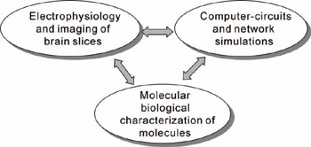
Figure 1
|
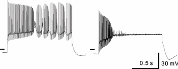
Figure 2. Firing pattern of the cerebellar Purkinje cells.
Normal Purkinje cells exhibit a bursting pattern of action potential generation (left), but Purkunje cells from ataxic rolling mice show only an abortive firing (right).
|
Staff
 |
Professor:
IMOTO, Keiji, MD, PhD
Graduated from Kyoto University Faculty of Medicine. Medical Staff, National Utano Hospital. Instructor, Lecturer, and Associate Professor, Kyoto University Faculty of Medicine. Research Associate, Max-Planck-Institut für medizinische Forschung. 1995 Professor, NIPS.
Specialty: Molecular and cellular neurophysiology |
 |
Associate Professor:
MIYATA, Mariko, MD, PhD
Graduated from Tokyo Women's Medical University Graduate School. Research Scientist in Frontier Research System, RIKEN. Research associate in Tokyo Women's Medical University. 2002 Associate Professor, NIPS.
Specialty: Neurophysiology |
 |
Assistant Professor:
YAMAGATA, Yoko, MD, PhD
Graduated from Kyoto University Graduate School of Medicine. Research Associate, Kyoto University Faculty of Medicine. Postdoctoral Fellow, The Rockefeller University. 1991 Research Associate, NIPS.
Specialty: Biochemistry, Neurochemistry |
 |
Assistant Professor:
SATAKE, Shinichiro, PhD
Graduated from Nagoya University Graduate School of Science. Postdoctoral Fellow of Mitsubishi Kagaku Institute of Life Sciences, Research Fellow of CREST (JST). 2002 Research Associate, NIPS.
Specialty: Neurophysiology, Neurochemistry
|
 |
Assistant Professor:
INOUE, Tsuyoshi, PhD
Graduated from University of Tokyo Graduate School of Pharmaceutical Sciences. Postdoctoral Fellow of Case Western Reserve University. 2003 Research Fellow, NIPS. 2003 Research Associate, NIPS.
Specialty: Neurophysiology |
 |
Postdoctoral Fellow:
SASAKI, Sachie, PhD
Graduated from the Graduate University for Advanced Studies. 2004 Research Fellow, NIPS.
Specialty: Neurophysiology |
 |
Postdoctoral Fellow:
KODAMA, Takashi
Graduated from the Graduate University for Advanced Studies. 2006 Research Fellow, NIPS.
Specialty: Neurophysiology |
|