|
|
|
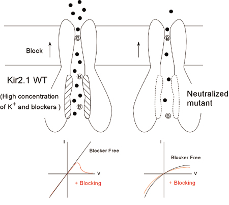
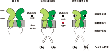
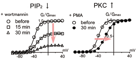
DEPARTMENT OF MOLECULAR PHYSIOLOGYDivision of Biophysics and NeurobiologyIon channels, receptors and G proteins play critical roles for the excitability and its regulation of neurons. We focus on these molecules which enable brain function. From the biophysical point of view, we study structure-function relationships, regulation mechanisms and dynamic structural rearrangements of ion channels and receptors. We also plan to study the functional significance of specific features of ion channels and receptors in the brain function by making knock-in mice and by studying their abnormalities in the synaptic transmission and whole animal behavior. Specific themes of research projects currently running are as follows.
|
|||||||||||||||||||||||

Staff |
|
 |
Professor: KUBO, Yoshihiro, MD, PhD 1985 Graduated from University of Tokyo, Faculty of Medicine. 1989 Completed the doctoral course in Medical Science, University of Tokyo. 1989-2000 Researcher, Tokyo Metropolitan Institute for Neuroscience. (1991-1993: Post-doc, University of California, San Francisco). 2000 Professor, Tokyo Medical and Dental University Graduate School of Medicine. 2003 Professor, NIPS. Specialty: Biophysics, Neurobiology |
 |
Associate Professor: TATEYAMA, Michihiro, PhD 1990 Graduated from University of Tokyo, Faculty of Pharmacology. 1995 Completed the doctoral course in Pharmacology, University of Tokyo. 1995-2000 Assistant Professor, Juntendo University School of Medicine. 2000-2002 Research Fellow, Columbia University. 2002-2004 Research Fellow, CREST. 2004 Associate Professor, NIPS. Specialty: Pharmacology, Physiology |
 |
Assistant Professor: NAKAJO, Koichi, PhD 1997 Graduated from University of Tokyo, College of Arts and Sciences. 2002 Completed the doctoral course in Life Science, University of Tokyo Graduate School of Arts and Sciences. 2002 Inoue Research Fellow. 2004 Research Fellow, NIPS. 2005 Assistant Professor, NIPS. Specialty: Molecular and Cellular Physiology |
 |
Postdoctoral Fellow: ITOH, Masayuki, PhD 2001 Graduated from Toho University, Faculty of Science. 2006 Completed the doctoral course in Science, Toho University. 2006 Research Fellow, NIPS. Specialty: Molecular biology |
Division of Neurobiology and BioinformaticsDuring the course of formation of the mammalian central nervous system, neuroepithelial cells differentiate into various kinds of cells to make a fine three-dimensional network. Our goal is to understand genetic control over these processes. As a first step, we have cloned several genes that are specifically expressed in a certain type of brain cells and are investigating their role on cell fate determination. Neural cells are known to leave the ventricular zone after their commitment, and migrate towards destinations. While radial neuronal migration has been studied extensively in the developing cerebral and cerebellar cortices, mechanisms underlining tangential migration of neuronal and glial progenitors remains unclear. We are employing in ovo or in utero electroporation method to introduce exogenous genes in developing central nervous system, and studying mode and mechanisms of neural cell migration. We are making use of hereditary mutant mice that exhibit abnormal development of the nervous system. We also use in situ hybridization and immunohistochemical technique to study cell lineages during development of the nervous system. Neural stem cells, which are ultimate lineage precursors to all neurons and glia in the mammalian brain, are present not only in embryonic but also in adult brains, and contribute to adult neurogenesis. We are investigating molecular mechanisms underlying the generation, proliferation, maintenance, differentiation, and senescence of the neural stem cells, which will clarify their in vivo kinetics and function. An automated system to analyze N-linked sugar chains was developed to study their biological roles during development and tumorigenesis. New retroviral vectors are also constructed for efficient gene delivery, which will be used for cancer gene therapy.
|
|
 |
 |
|
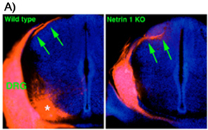
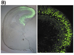
A) Aberrant projection of DRG axons to the dorsal spinal cord in the Netrin 1 deficient mouse. DRG axons are labeled by DiI application to DRG. In the wild type spinal cord, DRG fibers form axon bundle in the dorsolateral superficial part (arrows in left picture). By contrast, DRG axons in the Netrin 1 deficient mouse spinal cord enter the mantle layer directly and form aberrant axon bundle within it (arrows in right picture). Asterisk in left picture indicates motoneurons labeled retrogradely, which is not a defect of Netrin deficiency. B) In utero electroporation was carried out for plasmid DNA transfer. Green fluorescent protein (GFP) expression vector was injected into lateral ventricle and electroporated in utero. The cells in the restricted region were observed to express GFP | |
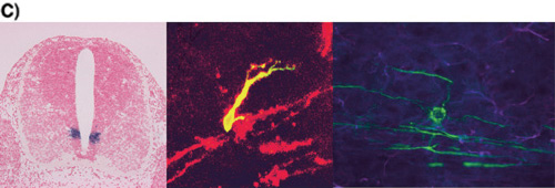
C) Oligodendrocyte development.(Left) pMN domain which is the site of oligodendrogenesis. Expression of Olig2 gene in embryonic day 12 spinal cord. Olig2 (purple) is expressed ventral ventricular zone called pMN domain.(Middle) Migrating oligodendrocyte progenitor. Oligodendrocyte progenitor is double-stained by anti-GFP antibody (green) and O4 antibody (red). O4 is an oligodendrocyte lineage specific marker.(Right) Myelinating oligodendrocyte Mature oligodendrocyte (green) is observed with extending processes toward several axons. |
Staff |
|
 |
Professor: IKENAKA, Kazuhiro, PhD 1975 Graduated from Faculty of Science, Osaka University. 1980 Graduated from the doctoral course at Osaka University, PhD. 1980 Instructor at Institute for Protein Research, Osaka University. 1991 Associate Professor at Institute for protein Research, Osaka University. 1992 Professor, NIPS. Specialty: Molecular Neurobiology |
 |
Associate Professor: ONO, Katsuhiko, PhD 1980 Graduated from Faculty of Science, Okayama University. 1982 Graduated from the master course at Okayama University. 1988 PhD from Okayama University Medical School. 1982 Research Associate at Okayama University Medical School, 1993 Assistant professor at Okayama University Medical School. 1995 Associate professor at Shimane Medical University. 2003 Associate professor at NIPS. Specialty: Neural Development |
 |
Associate Professor: HITOSHI, Seiji, MD, PhD 1988 Graduated from Faculty of Medicine, University of Tokyo. MD. 1993 Board-certified neurologist by Japanese Society for Neurology. 1997 PhD from Graduate School of Medicine, University of Tokyo. 1997 Special Postdoctoral Researcher at the Institute of Physical and Chemical Research (RIKEN). 1999 Postdoctoral Fellow at University of Toronto. 2003 Assistant Professor at University of Tokyo. 2003 Associate Professor at NIPS. Specialty: Developmental Neurobiology, Neurology |
 |
Assistant Professor: TAKEBAYASHI, Hirohide, MD, PhD 1995 Graduated from Kyoto University, Faculty of Medicine. 1999 Graduated from Kyoto University, Graduate School of Medicine.1999 Postdoctoral Fellow, Kyoto University, 2002 Research Associate, NIPS. Specialty: Molecular Neurobiology |
 |
Assistant Professor: TANAKA, Kenji, MD, PhD 1997 Graduated from Keio University, School of Medicine. 1997-1999 Resident in Department of Neuropsychiatry, Keio University, School of Medicine. 2003 Completed the doctoral course in Keio University. 2003 Research Associate, NIPS. 2004 Assistant Professor, NIPS. Specialty: Neurochemistry, Biological psychiatry |
 |
Postdoctoral Fellow: DING, Lei, MD, PhD 1990 Graduated from DaLian Medical University ,China. 2003 Completed doctoral course in medicine at the Hokkaido University. 2003 Research Fellow, NIPS. 2004 Postdoctoral Fellow. Specialty: Neural Development and Neuroanatomy |
 |
Postdoctoral Fellow: NARUSE, Masae, PhD 2001 Graduated from Tokyo Institute of Technology, Department of Bioscience and Biotechnology. 2003 Graduated from the master course in Tokyo Institute of Technology, Department of Bioscience and Biotechnology. 2006 Graduated from the doctoral course in Life Science, the Graduate University for Advanced Studies, PhD. 2006 Postdoctoral Fellow. Specialty: Neural Development |
 |
Postdoctoral Fellow: HIGASHI, Mikito, PhD 2002 Graduated from Department of Bio-Medical Engineering, Tokai University. 2006 Graduate from the doctoral course at Graduate University for Advanced Studies, PhD. 2006 Postdoctoral Fellow, NIPS. Specialty: Molecular Neurobiology, Stem Cell Biology |
 |
Postdoctoral Fellow: WATANABE, Keisuke, PhD 2001 Graduated from Faculty of Agriculture, Tohoku University. 2003 Graduated from the master course in Agricultural Science, Tohoku University. 2006 Graduated from the doctoral course in Life Science, the Graduate University for Advanced Studies, PhD. 2006 Postdoctoral Fellow, NIPS. Specialty: Neural Development |
Division of Intracellular MetabolismCell signaling that generates proper cell responses to various stimuli is the essence of life. To understand its mechanism is one of the goals of life sciences. This division is aiming to elucidate the spatio-temporal regulation mechanisms underlying cell signaling, focusing on the dynamics of ion channels, cytoskeletons, and adhesion molecules by use of electrophysiological and advanced imaging techniques. The subjects of research are, (1) Cell signaling in response to mechanical stimuli: (2) Intracellular Ca2+ signaling: (3) Proton signaling: |
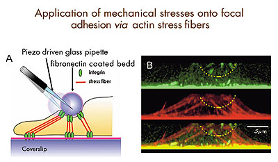
Fig.1: Diagram for mechanical stimulation of focal adhesions through stress fibers. Left: A fibronectin-coated glass bead connected to the basal focal adhesions via stress fibers. By displacing the bead, we can apply localized mechanical stimuli onto focal adhesions, while recording the surface dynamics of intracellular calcium and integrin by near field microscopy. Right: Projected side views of focal adhesions (top, green spots), stress fibers (middle, red strands), and their superimposition (bottom) in an endothelial cell. |
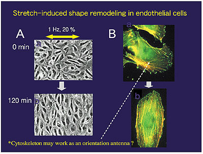
Fig.2: Stretch-induced shape remodeling. Left: When subjected to uniaxial cyclic stretch, endothelial cells cultured on an elastic silicone membrane change their shape from cobble stone-like to spindle-like by aligning their long axis perpendicular to the stretch axis. Right: Dynamic rearrangement of focal adhesions (green spots) and stress fibers (orange strands) before (top) and after (bottom) remodeling. |
Staff |
|
 |
Professor: SOKABE, Masahiro, PhD 1973 Graduated from Osaka University, Faculty of Engineering Siences. 1975 Completed a master course in Physics, Osaka University. 1975 Research Associate, Osaka University, Faculty of Human Sciences. 1985 Lecturer. 1987 Associate Professor. 1992 Professor, Nagoya University School of Medicine, Department of Physiology. 1999 Professor, Nagoya University Graduate School of Medicine, Department of Cell Science. 2003 Adjunct Professor, NIPS. Speciality: Ion Channel and Cell Biophysics, Neuroscience |
 |
Associate Professor: KUNO, Miyuki, PhD 1979 Graduated from Osaka City University School of Medicine. 1981 Research Associate, Osaka City University. 1984 PhD degree in Medicine, Osaka City University. 1986 Lecturer. 1992 Associate Professor. 2000 Associate Professor, Osaka City University Graduate School of Medicine, Department of Molecular and Cellular Physiology. 2004 Adjunct Associate Professor, NIPS. Speciality: Ion Channel and Cell Physiology |
 |
Assistant Professor: MOHRI, Tatsuma, PhD 1978 Graduated from Yamaguchi University. 1981 Completed a master course in Physics, Kanazawa University. 1991 Completed a doctoral course in Life Chemistry, Tokyo Institute of Technology. 1991 Jean and Katsuma Dan Fellow, Hopkins Marine Station Stanford University. 1991 Postdoctoral Associate and 1993 Research Associate, University of Miami School of Medicine. 1995 Postdoctoral Researcher, University of California Davis. 1996 Research Associate, NIPS. Speciality: Cell Biology, Cell Physiology |
 |
Postdoctoral Fellow: HIRATA, Hiroaki, PhD 1998 Graduated from Tohoku University, Faculty of Science. 2000 Completed a master course in Physics, Tohoku University. 2003 Completed a doctoral course in Physics, Tohoku University. May 2003 Research Fellow, JST. Nov 2003 Research Fellow, NIPS. Specialty: Cell Biophysics |
|
|
|
| Copyright(C) 2006 NIPS (National Institute for Physiological Sciences) | |