CENTER FOR BRAIN EXPERIMENT
|
The ultimate object of this laboratory is to clarify the relations between structures and functions in the brain. To study them, a high voltage electron microscope (H-1250M) which is specially designed for biological and medical research is available since 1982. The daily accelerating voltage of the microscope is 1,000kV. The pressure near the specimen position is less than 7×10⁻⁶Pa and the magnification ranges 1k to 1,000k times. Transmission images of thick specimens till about 5mm can be taken.
Since this is the only high voltage electron microscope in the biological field in Japan, the collaborative programs are carried out by about 15 research groups from Universities etc. on three projects: 1) Three-dimensional analysis of fine structures of biological specimens 2) High resolution observation of biological specimens 3) Observation of biological specimens in their natural conditions.
Facilities for tissue culture and light microscopy are also provided. Cryostats, microtomes and fluorescence microscopes with a high-resolution colour cooled CCD camera system are prepared for immunohistochemistry. Inverted microscopes with a time lapse video system are prepared to observe cultured cells.
|
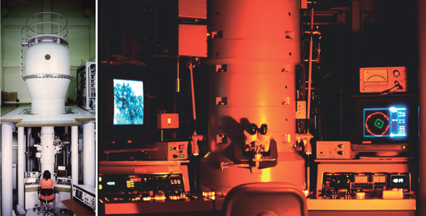
High voltage electron microscope (H-1250M: 1,000kV)
Specially designed for the exclucive use of medical and biological specimens
|
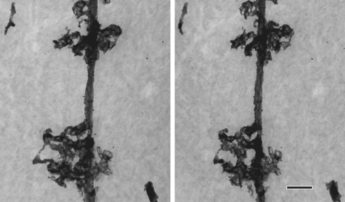
Cell processes of a Bergmann glia in the rat cerebellum revealed by Golgi staining
Stereo images taken at ±8º tilt at 1,000kV. Specimen thickness: 3mm. Scale bar: 2mm.
|
Staff
 |
Associate Professor:
ARII, Tatsuo, PhD
1967 Graduated from Tohoku University, Faculty of Science. 1972 Completed the doctoral course in Engineering, Nagoya University. 1972 Research Associate, Nagoya University. 1973 Research Associate, Regensburg University. 1976 Research Associate, Nagoya University. 1979 Associate Professor, NIPS.
Speciality: Electron Microscopy |
 |
Assistant Professor:
FURUYA, Sonoko, PhD
1970 Graduated from University of Tokyo, Faculty of Pharmacy. 1975 Completed the doctoral course in Pharmacy, University of Tokyo. 1975 Research Associate, Nihon Medical College. 1978 Research Associate, NIPS.
Speciality: Tissue Culture and Histology
|
In order to investigate the brain mechanism underlying our mental ability such as cognition, voluntary movement, thinking, or will, we have to experiment on the human brain. Some non-invasive techniques for measuring brain are certainly useful for the purpose. However, they are still insufficient in the quality of information. To overcome the limitations, researches on the brain are carried out here in both the human and monkey subjects using various techniques including direct recordings of cortical field potential, magnetoencephalography, and positron emission tomography.
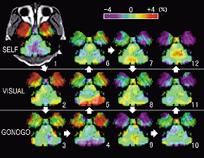
Figure 1. Brain activity during three kinds of movement tasks (a specimen record in one day).
The monkey was engaged in hand movement tasks to get a reward in three different sets of circumstances; i.e. the self-initiated movement task (SELF), the visually-initiated movement task (VISUAL), and the color-discriminating go/no-go task with asymmetrical reinforcement (GONOGO). Arrows and numbers indicate the order in which they were taken. Although the activity fluctuates, a statistical analysis across repetitive measurements can extract the significant activity changes (Figures 2, 3, and 4)
|
|
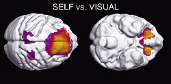
Figure 2. Regions of activated in SELF task.
|
|
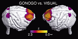
Figure 3. Regions activated in GONOGO task.
|
|
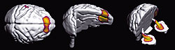
Figure 4. Regions of significant decrease in activity during task repetition in a day.
The decline in the activity of the limbic and prefrontal regions may be a reflection of a decline in the willingness.
|
Staff
 |
Associate Professor:
TSUJIMOTO, Toru, MD, PhD
1986 Graduated from Kyoto University, Faculty of Medicine. 1990 Completed the doctoral course in Medicine, Kyoto University. 1993 Research Associate, NIPS. 1994 Research Associate, Kyoto University. 1999 Associate Professor, NIPS.
Speciality: Neurophysiology
|
i) Two-photon Microscopy Imaging Group
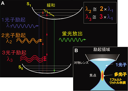
In order to support international or domestic collaborative researches, we have developed the most advanced "two-photon microscopy". The microscopy has given us important insights into secretory functions in neural and secretory gland cells. We have especially proved the existence of "sequential compound exocytosis". We explore two-photon microscopy by incorporation of molecular biology, patch-clamp, photo-activated probes, and non-linear optics techniques. The final goal is to reveal "missing-links" underlying between molecular, cellular, and physiological functions of human bodies (Figs. 1- 3).
ii) Computer & Network group
Computing and network support are indispensable for research activity. In this section, we have a HP ES45 system for data analyses and simulation and two technical staffs support high-speed and reliable network and peripheral device for in-house information service, high-quality digital color printing, etc. Technological developments for the best use of these facilities are also underway (Figs. 4, 5).
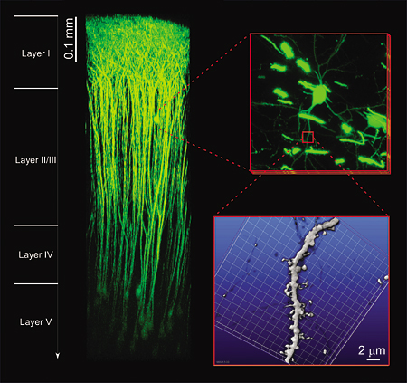 |
| Figure 1. "in vivo" two-photon microscopy. The superior tissue penetration of our newly constructed "in vivo" two-photon microscopy can visualize neural activities in a living brain. EYFP fluorescence can be detected from deeper layers than 0.9 mm beneath the surface in an anaesthetized mouse. 3D-reconstruction of individual neural cells spreading in the layers I-V is available without any degradation of the spatial resolution of sub-micron. (Collaborative research with Prof. J. Nabekura) |
 |
| Figure 2. Multi-photon excitation process. By using near infrared femto-second laser, multi-photon excitation of molecules can be elicited by simultaneous absorption of photons (A) at the focal point of an objective lens (B). Two-photon excitation imaging (two-photon microscopy) has deeper tissue penetration, little out-of-focal light absorption and least phototoxic effects. It is thus quite suitable for investigating molecular and cellular events within thick intact tissues. Moreover, it allows simultaneous multi-color imaging and fluorescence correlation measurement. The fusion pore opening and its dynamics can be resolved of a nanometer order (Nature Cell. Biol., 3: 253, 2001, Science, 297:1349, 2002). |
 |
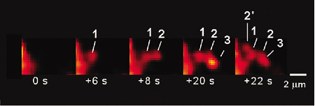
| Figure 3. The discovery of "sequential compound exocytosis". Two-photon microscopy has demonstrated sequential progression of exocytosis deep into the cytosol in exocrine glands. Such sequential compound exocytosis is now considered as used commonly for physiological secretions in a wide variety of cells and organs (Nature Cell. Biol., 3:253, 2001, EMBO J, 25:673, 2006). |
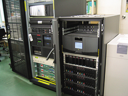
Figure 4. Computer System for Data Analysis in Physiology
|
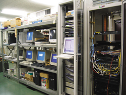
Figure 5. Network servers
|
Staff
 |
Associate Professor:
NEMOTO, Tomomi, PhD
1991 Graduated from, Department of Physics, Faculty of Science, the University of Tokyo. 1996 Completed the doctoral course in Applied Physics in Tokyo Institute of Technology. 1996-1997 Frontier Researcher and Special Postdoctoral Researcher, RIKEN. 1997-1999 Research fellow, the University of Tokyo. 1999-2005, Assistant professor, NIPS. 2001-2004, Researcher, PRESTO, JST.
Specialty: Cell physiology, Biophysics.
|
|
|