DEPARTMENT OF MOLECULAR PHYSIOLOGY
Ion channels, receptors and G proteins play critical roles for the excitability and its regulation of neurons. We focus on these molecules which enable brain function. From the biophysical point of view, we study structure-function relationships, regulation mechanisms and dynamic structural rearrangements of ion channels and receptors. We also plan to study the functional significance of specific features of ion channels and receptors in the brain function by making knock-in mice and by studying their abnormalities in the synaptic transmission and whole animal behavior. Specific themes of research projects currently running are as follows.
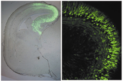
- Structure-function relationship of inwardly rectifying K+ channels.
- Molecular mechanisms and functional significance of the Ca2+/Gd3+ sensing function of metabotropic glutamate receptor.
- Analysis of the dynamic structural rearrangements of ion channels and receptors by FRET measurement under evanescent field illumination.
- Molecular mechanisms of the regulation of M- channel function by muscarinic stimulation.
- Changes of the local environment and movement of S4 domain of KCNQ1 channel by assembled KCNE subunit.
- Voltage-dependent gating and expression density dependent changes of the pore properties of ATP receptor channel P2X2.
- Molecular identification and functional analysis of Prestin complex, a motor protein of the outer hair cell.
- cDNA cloning and functional analysis of RGS family, regulators of G protein signaling.
- Purification of recombinant proteins of ATP receptor channel P2X2 toward single particle structure analysis.
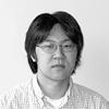
Fig. 1. Functional roles of charged amino acid residues on the wall of the cytoplasmic pore of inward rectifier K+ channel Kir2.1. (Fujiwara and Kubo, J. Gen. Physiol., 2006)
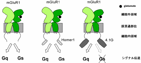
Fig. 2. Coupling profile of the metabotropic glutamate receptor 1a is regulated by the C-terminal domain. (Tateyama and Kubo, Mol. Cell. Neurosci., 2007)
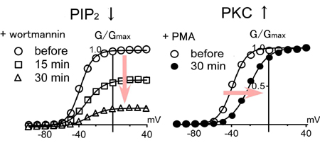
Fig. 3. Different roles of PIP2 and PKC during muscarinic inhibition of KCNQ/M channels. (Nakajo and Kubo, J. Physiol., 2005)
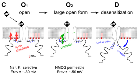
Fig. 4. Regulation of the desensitization and ion selectivity of ATP-gated P2X2 channels by phosphoinositides. (Fujiwara and Kubo, J. Physiol., 2006)
Staff
 |
Professor:
KUBO, Yoshihiro, MD, PhD
1985 Graduated from University of Tokyo, Faculty of Medicine. 1989 Completed the doctoral course in Medical Science, University of Tokyo. 1989-2000 Researcher, Tokyo Metropolitan Institute for Neuroscience. (1991-1993: Post-doc, University of California, San Francisco). 2000 Professor, Tokyo Medical and Dental University Graduate School of Medicine. 2003 Professor, NIPS.
Specialty :Biophysics, Neurobiology |
 |
Associate Professor:
TATEYAMA, Michihiro, PhD
1990 Graduated from University of Tokyo, Faculty of Pharmacology. 1995 Completed the doctoral course in Pharmacology, University of Tokyo. 1995-2000 Assistant Professor, Juntendo University School of Medicine. 2000-2002 Research Fellow, Columbia University. 2002-2004 Research Fellow, CREST. 2004 Associate Professor, NIPS.
Specialty: Pharmacology, Physiology |
 |
Assistant Professor:
NAKAJO, Koichi, PhD
1997 Graduated from University of Tokyo, College of Arts and Sciences. 2002 Completed the doctoral course in Life Science, University of Tokyo Graduate School of Arts and Sciences. 2002 Inoue Research Fellow. 2004 Research Fellow, NIPS. 2005 Assistant Professor, NIPS.
Specialty: Molecular and Cellular Physiology |
 |
Postdoctoral Fellow:
ITOH, Masayuki, PhD
2001 Graduated from Toho University, Faculty of Science. 2006 Completed the doctoral course in Science, Toho University. 2006 Research Fellow, NIPS.
Specialty: Molecular biology |
During the course of formation of the mammalian central nervous system, neuroepithelial cells differentiate into various kinds of cells to make a fine three-dimensional network. Our goal is to understand genetic control over these processes. As a first step, we have cloned several genes that are specifically expressed in a certain type of brain cells and are investigating their role on cell fate determination. Neural cells are known to leave the ventricular zone after their commitment, and migrate towards destinations. While radial neuronal migration has been studied extensively in the developing cerebral and cerebellar cortices, mechanisms underlining tangential migration of neuronal and glial progenitors remains unclear. We are employing in ovo or in utero electroporation method to introduce exogenous genes in developing central nervous system, and studying mode and mechanisms of neural cell migration.
We are making use of hereditary mutant mice that exhibit abnormal development of the nervous system. We also use in situ hybridization and immunohistochemical technique to study cell lineages during development of the nervous system.
Neural stem cells, which are ultimate lineage precursors to all neurons and glia in the mammalian brain, are present not only in embryonic but also in adult brains, and contribute to adult neurogenesis. We are investigating molecular mechanisms underlying the generation, proliferation, maintenance, differentiation, and senescence of the neural stem cells, which will clarify their in vivo kinetics and function.
An automated system to analyze N-linked sugar chains was developed to study their biological roles during development and tumorigenesis.
New retroviral vectors are also constructed for efficient gene delivery, which will be used for cancer gene therapy.
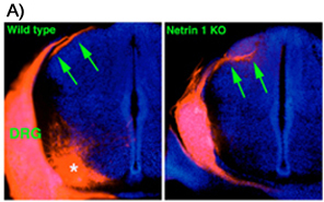
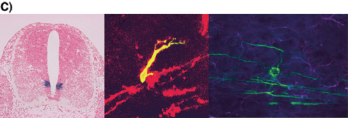
A) Aberrant projection of DRG axons to the dorsal spinal cord in the Netrin 1 deficient mouse. DRG axons are labeled by DiI application to DRG. In the wild type spinal cord, DRG fibers form axon bundle in the dorsolateral superficial part (arrows in left picture). By contrast, DRG axons in the Netrin 1 deficient mouse spinal cord enter the mantle layer directly and form aberrant axon bundle within it (arrows in right picture). Asterisk in left picture indicates motoneurons labeled retrogradely, which is not a defect of Netrin deficiency.
B) In utero electroporation was carried out for plasmid DNA transfer. Green fluorescent protein (GFP) expression vector was injected into lateral ventricle and electroporated in utero. The cells in the restricted region were observed to express GFP
C) Oligodendrocyte development.(Left) pMN domain which is the site of oligodendrogenesis. Expression of Olig2 gene in embryonic day 12 spinal cord. Olig2 (purple) is expressed ventral ventricular zone called pMN domain.(Middle) Migrating oligodendrocyte progenitor. Oligodendrocyte progenitor is double-stained by anti-GFP antibody (green) and O4 antibody (red). O4 is an oligodendrocyte lineage specific marker.(Right) Myelinating oligodendrocyte Mature oligodendrocyte (green) is observed with extending processes toward several axons.
Staff
 |
Professor:
IKENAKA, Kazuhiro, PhD
1975 Graduated from Faculty of Science, Osaka University. 1980 Graduated from the doctoral courseat Osaka University, PhD. 1980 Instructor at Institute for Protein Research, Osaka University. 1991 Associate Professor at Institute for Protein Research, Osaka University. 1992 Professor, NIPS.
Specialty: Molecular Neurobiology |
 |
Associate Professor:
ONO, Katsuhiko, PhD
1980 Graduated from Faculty of Science, Okayama University. 1982 Graduated from the master course at Okayama University. 1988 PhD from Okayama University Medical School. 1982 Research Associate at Okayama University Medical School, 1993 Assistant professor at Okayama University Medical School. 1995 Associate professor at Shimane Medical University. 2003 Associate professor at NIPS.
Specialty: Neural Development |
 |
Associate Professor:
HITOSHI, Seiji, MD, PhD
1988 Graduated from Faculty of Medicine, University of Tokyo. MD. 1993 Board-certified neurologist by Japanese Society for Neurology. 1997 PhD from Graduate School of Medicine, University of Tokyo. 1997 Special Postdoctoral Researcher at the Institute of Physical and Chemical Research (RIKEN). 1999 Postdoctoral Fellow at University of Toronto. 2003 Assistant Professor at University of Tokyo. 2003 Associate Professor at NIPS.
Specialty: Developmental Neurobiology, Neurology |
 |
Assistant Professor:
TAKEBAYASHI, Hirohide, MD, PhD
1995 Graduated from Kyoto University, Faculty of Medicine. 1999 Graduated from Kyoto University, Graduate School of Medicine. 1999 Postdoctoral Fellow, Kyoto University, 2002 Research Associate, NIPS.
Specialty: Molecular Neurobiology |
 |
Assistant Professor:
TANAKA, Kenji, MD, PhD
1997 Graduated from Keio University, School of Medicine. 1997-1999 Resident in Department of Neuropsychiatry, Keio University, School of Medicine. 2003 Completed the doctoral course in Keio University. 2003 Research Associate, NIPS. 2004 Assistant Professor, NIPS.
Specialty: Neurochemistry, Biological psychiatry |
 |
Postdoctoral Fellow:
GOTOH, Hitoshi, PhD
2002 Graduated from Kobe University, Faculty of Agriculture. 2004 Graduated from the master course in Osaka University, Faculty of Science. 2007 Graduated from the doctoral course in Osaka University, Faculty of Science, PhD. 2007 Postdoctoral Fellow, NIPS.
Specialty: Molecular Neurobiology |
 |
Postdoctoral Fellow:
NARUSE, Masae, PhD
2001 Graduated from Tokyo Institute of Technology, Department of Bioscience and Biotechnology. 2003 Graduated from the master course in Tokyo Institute of Technology, Department of Bioscience and Biotechnology. 2006 Graduated from the doctoral course in Life Science, the Graduate University for Advanced Studies, PhD. 2006 Postdoctoral Fellow.
Specialty: Neural Development |
Cell signaling that generates proper cell responses to various stimuli is the essence of life. To understand its mechanism is one of the goals of life sciences. This division is aiming to elucidate the spatio-temporal regulation mechanisms underlying cell signaling, focusing on the dynamics of ion channels, cytoskeletons, and adhesion molecules by use of electrophysiological and advanced imaging techniques.
The subjects of research are,
(1) Cell signaling in response to mechanical stimuli:
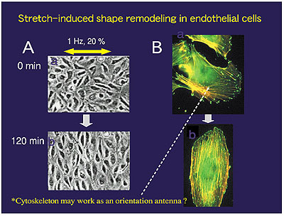
Virtually every cell can properly respond to mechanical stimuli, e.g., electrical responses in the inner ear hair cells and cutaneous mechanoreceptors, disuse atrophy in muscle and bone under microgravity, or shear stress induced NO production in endothelial cells. However, its molecular mechanisms are largely unknown due to the ambiguity of the mechanotransduction process in cells. We therefore focus on SA channels and the cytoskeleton/focal adhesion complex as representative cell mechanosensors and investigate their roles in mechanosignaling through the development of innovative light microscopy and micro mechanical manipulation of the cell (Fig.1). A typical subject is stretch-induced shape remodeling, where endothelial cells align their long axis perpendicular to the stretch axis. This response includes many intriguing functions, such as sensation of force direction and spatio-temporal integration of dynamics of stress fibers and focal adhesions during their rearrangement (Fig.2).
(2) Intracellular Ca2+ signaling:
When various mechanical stimuli, such as have induced by cell-cell interaction or stimuli induced by biological activators such as hormones, are given to a cell, the cell exhibits intracellular Ca2+ changes in response to them. The Ca2+ changes are modulated and processed on to the next signal pathways, leading to various significant cell functions. The process called intracellular Ca2+ signaling is one of the most significant and major signal transduction mechanisms in cells of almost all organisms. We use Ca2+ and Na+ imaging techniques, and electrophysiological methods to perform our experiments in addition to cellular manipulations such as microsurgery and microinjection of materials into cells. We presently focus on the Ca2+ signaling in stretched-induced or migrating cells to investigate the mechanisms aforementioned in (1). We also investigate the mechanisms of fertilization and oocyte maturation in mammals through the study of the Ca2+ oscillations and Ca2+ increase.
(3) Proton signaling:
Hydrogen ion (proton, H+) is an important signaling ion that determines pH and participates in a variety o Hydrogen ion (proton, H+) is an important signaling ion that determines pH and participates in a variety of biological responses, for instance bone remodeling, natural immunity, and pain sensation. H+-transferring molecules at the plasma membrane serve to regulate the pH environment dynamically. Voltage-gated H+ channels function as sensitive pH monitors and acid-secreting apparatuses, and have been cast as a key player in the processes of H+ signaling. The primary goal of this study is employing H+ channels to elucidate the mechanisms underlying H+ mobilization linked with cellular functions.
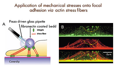
Fig. 1:Diagram for mechanical stimulation of focal adhesions through stress fibers. Left: A fibronectin-coated glass bead connected to the basal focal adhesions via stress fibers. By displacing the bead, we can apply localized mechanical stimuli onto focal adhesions, while recording the surface dynamics of intracellular calcium and integrin by near field microscopy. Right: Projected side views of focal adhesions (top, green spots), stress fibers (middle, red strands), and their superimposition (bottom) in an endothelial cell.
Fig. 2:Stretch-induced shape remodeling. Left: When subjected to uniaxial cyclic stretch, endothelial cells cultured on an elastic silicone membrane change their shape from cobble stone-like to spindle-like by aligning their long axis perpendicular to the stretch axis. Right: Dynamic rearrangement of focal adhesions (green spots) and stress fibers (orange strands) before (top) and after (bottom) remodeling.
Staff
 |
Professor:
SOKABE, Masahiro, PhD
1973 Graduated from Osaka University, Faculty of Engineering Siences. 1975 Completed a master course in Physics, Osaka University. 1975 Research Associate, Osaka University, Faculty of Human Sciences. 1985 Lecturer. 1987 Associate Professor. 1992 Professor, Nagoya University School of Medicine, Department of Physiology. 1999 Professor, Nagoya University Graduate School of Medicine, Department of Cell Science. 2003 Adjunct Professor, NIPS.
Speciality: Ion Channel and Cell Biophysics, Neuroscience |
 |
Associate Professor:
KUNO, Miyuki, PhD
1979 Graduated from Osaka City University School of Medicine. 1981 Research Associate, Osaka City University. 1984 PhD degree in Medicine, Osaka City University. 1986 Lecturer. 1992 Associate Professor. 2000 Associate Professor, Osaka City University Graduate School of Medicine, Department of Molecular and Cellular Physiology. 2004 Adjunct Associate Professor, NIPS.
Speciality: Ion Channel and Cell Physiology |
 |
Assistant Professor:
MOHRI, Tatsuma, PhD
1978 Graduated from Yamaguchi University. 1981 Completed a master course in Physics, Kanazawa University. 1991 Completed a doctoral course in Life Chemistry, Tokyo Institute of Technology. 1991 Jean and Katsuma Dan Fellow, Hopkins Marine Station Stanford University. 1991 Postdoctoral Associate and 1993 Research Associate, University of Miami School of Medicine. 1995 Postdoctoral Researcher, University of California Davis. 1996 Research Associate, NIPS.
Specialty: Cell Biology, Cell Physiology |
 |
Postdoctoral Fellow:
HIRATA, Hiroaki, PhD
1998 Graduated from Tohoku University, Faculty of Science. 2000 Completed a master course in Physics, Tohoku University. 2003 Completed a doctoral course in Physics, Tohoku University. May 2003 Research Fellow, JST. Nov 2003 Research Fellow, NIPS.
Specialty: Cell Biophysics |
|
A novel methodology, when it is very informative, gives an aid to the opening of a novel scientific field. For example, MRI originally born from NMR in chemistry and primarily developed for diogonoes has outgrown to cover almost all medical sciences. We call such a productive innovation, emerged from an old regime but creative to a new field, as a strategic methodology. Integration of biosciences might bring about such a difficulty that a simple sum of constituent disciplines never makes a good start. Fusion of different disciplines can be encouraged by novel breakthroughs in methodology. The expected are new methods for three-dimensional structural analysis of single biological molecules and in situ functional observation of complex biological systems.
This laboratory works on methodological themes by relying on the technical breakthrough of imaging methods such as electron microscopy.
(1) Development and application of electron-phase microscopy: Different kinds of phase observation schemes have been developed including the novel optical principle for the reconstruction of complex wavefunctions. They are expected to enhance the contrast of biological samples which is inherently very poor in electron microscopy. Applications are:
i) direct visualization of protein molecules or cytoskeltons in the in vivo state of cells and tissues,
ii) structural and functional analyses of membrane proteins and viruses with the aid of single particle analysis.
(2) Biological transports: Transcellular and paracellurar mechanisms for transport of water, electrolytes, and substrates are investigatedby laying much emphasis on molecular mechanism of exocytosis and energy supply for transport in the exocrine glands.
(3) Aging process is, at least partially, regulated by the intracellular transport of neurotransmitter receptors and hormone receptors. We investigate the mechanism of receptor traffic especially via posttranslational modifications. Toward this goal, we developed mass microscope to visualize posttranslational modifications, and identified several novel enzymes for posttranslational modifications.
(4) Sorting in the endocytic pathway: The endocytic pathway functions as a sorting station for molecules that are destined either for lysosomes (a degrative pathway) or for recycling pathways, thereby determining the fate of endomembrane molecules. The physiological roles and the mechanisms of sorting in the endocytic pathway are investigated.
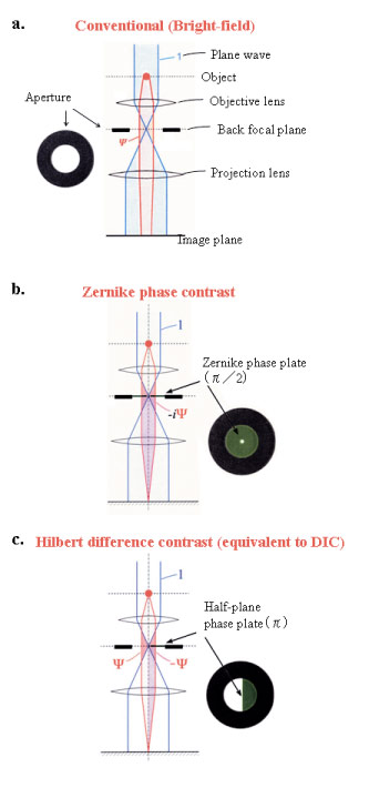
Fig. 1 Three kinds of schemes for electron-phase microscopy.
a. Conventional (bright-field) method to enhance the image contrast at the expense of a dehancement of the spatial resolution by defocusing.
b. Zernike phase contrast method to enhance the image contrast under the just focus condition by inserting a Zernike phase plate to the objective back-focal plane.
c. Hilbert differential method to obtain phase contrast images similar to light-microscopic DIC (Differential-Interference-Contrast) images by inserting a half-plane p phase plate to the objective back-focal plane.
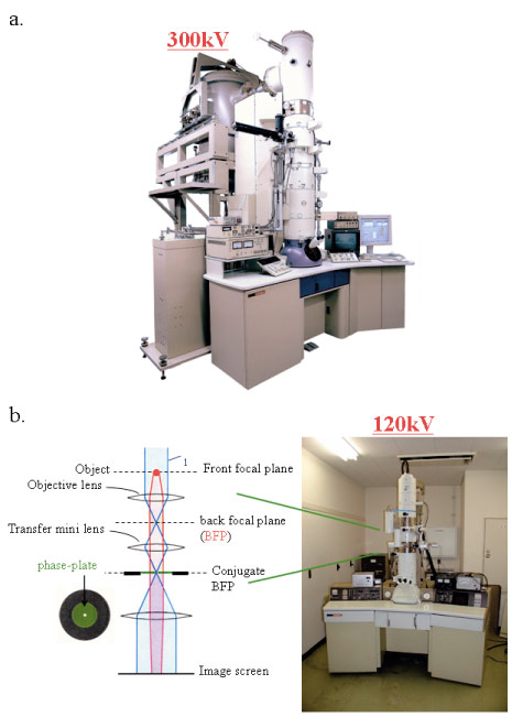
Fig. 2 Two kinds of electron-phase microscope models.
a. 300kV analytical cryo-electron microscope (equipped with a FEG, a He-stage and a w-filter) equipping phase plates at the back-focal plane.
b. 120kV TEM particularized to the phase contrast observation by furnishing a lens system immediately below the objective and fasillitating heating and precise positioning of phase plates.
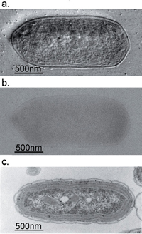
Fig. 3 Comparison of 300kV and 100kV TEM images for ice-embedded and plastic-embedded cyanobacterial cells.
a. A 300kV Hilbert differential image for an ice-embedded cyanobacterial whole cell, which holds a resolution sufficient for the identification of subcellular structures down to 2nm.
b. A 300kV conventional image shot for just the same sample as shown in a., of which low contrast makes it difficult to identify subcellular structures.
c. A 100kV conventional image for a plastic-embedded and thin-sectioned cyanobacterial cell, which was prepared with a chemical fixation, dehydration and a heavy metal staining. Due to the harsh and lengthy chemical treatments, subcellular structures are heavily damaged making their morphological preservation hard.
(Kaneko et al., J. Electro. Microsc. 54 (2005) 79)
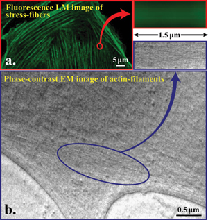
Fig. 4 Comparison of image resolving power between fluorescence light microscope and electron-phase microscope for actin filaments.
a. Fluorescence microscopic image of phalloidin stained stress fibers.
b. Hilbert differential TEM image (300kV) of actin filaments, of which bundles correspond to stress fibers.
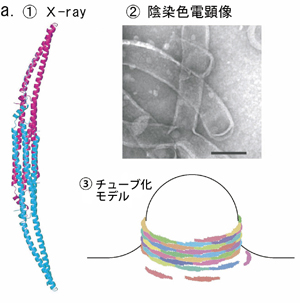
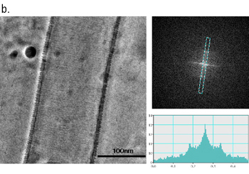
Fig. 5 Phase contrast cryo-TEM fills the gap beetweem X-ray crystallography and conventional TEM observation
a. Lipid interacting protein, PCH, has a domain called EFC-domain assumed to be responsible for the liposome tubulation. X-ray crystallography revealed a thread structure (①) and conventioned TEM observation with negative staining (②) showed a fixed size tubulation induced by the EFC-domain. From the two results a model as shown in ③ has been proposed.
b. The proposed model has been visually proven with the phase contrast cryo-TEM as shown in ①. The Fourier analysis (②,③) tells us the spacing of adjucently wound EFC-domain polymers being 4nm, which is just expected from the X-ray structure when modeled (Shimada et al., Cell, in press).
Staff
 |
Professor (concurrent, NIPS):
NAGAYAMA, Kuniaki, PhD
1968 Graduated from University of Tokyo. 1973 Completed the doctoral course in Science, University of Tokyo. 1974 Research Associate, University of Tokyo. 1984 Director, Biometrology Lab, JEOL Ltd. 1990 Project Leader, Nagayama Protein Array Project, ERATO, JRDC. 1993 Professor, The University of Tokyo. 1997 Professor, NIPS. 2001 Professor, Okazaki Institute for Integrative Bioscience (OIB).
Speciality: Biophysics, Electron Microscopy |
 |
Associate Professor (NIPS):
MURAKAMI, Masataka, MB, M.D.
1976 Graduated from Kyoto Prefectural University of Medicine. 1976 Research Associate, Osaka Medical College. 1981 Doctor of Medicine in Physiology of Osaka Medical College. 1983 Postdoctorial Fellow, Department of Physiology, University of Sydney. 1985 Associate Professor, NIPS. 2003 Associate Professor, OIB (Seconded from NIPS).
Speciality: Physiology of exocrine glands, Energy metabolism and transport of electrolyte and water, Paracellular Transport |
 |
Associate Professor:
SETOU, Mitsutoshi, MD, PhD
1994 Graduated from University of Tokyo, School of Medicine. 1998 Research Associate, University of Tokyo, School of Medicine. 2001 Doctor of Medicine in Cell biology and anatomy of University of Tokyo, School of Medicine. 2002 Principal Investigator of PRESTO, Japan Science and Technology Corporation. 2003 Associate Professor.
Speciality: Cell Biology and Anatomy, Intracellular transport, Receptor dynamics, Aging |
 |
Assistant Professor:
OHASHI, Masato, PhD
1986 Graduated from Kyoto University, Faculty of Science. 1992 Completed the doctoral course in Science, Kyoto University. 1992 Postdoctoral Fellow, Department of Neurobiology, University of Heidelberg. 1996 Assistant Professor, NIPS. 2003 Assistant Professor, OIB.
Speciality: Cell Biology |
 |
Assistant Professor:
DANEV, Radostin, PhD
1997 Graduated from Faculty of Physics, Sofia University, Sofia, Bulgaria. 2001 Completed the doctoral course in Science, The Graduate University for Advanced Studies, NIPS, Okazaki. 2001 Postdoctoral Fellow, NIPS. 2002 Research fellow, OIB. 2006 Assistant Professor, OIB.
Speciality: Solid State Physics, Electron Microscopy |
 |
Postdoctoral Fellow:
HAYASAKA, Takahiro, PhD
1997 Graduated from Shibaura Institute of Technology. 2003 Completed the doctoral course in Functional Control Systems. 2003 Postdoctoral Fellow, JST. 2004 Postdoctoral Fellow, Mitsubishi Kagaku Institute of Life Sciences. 2004 Postdoctoral Fellow, OIB.
Speciality: Biochemistry |
 |
Postdoctoral Fellow:
SHIGEMATSU, Hideki,
1994 Graduated from Tokyo Institute of Technology. 1999 Completed the doctoral course in Biotechnology, Tokyo Institute of Technology. 1999 Postdoctral Fellow, NIBH, 2000 Postdoctral Fellow, Kirin Brewery Co., Ltd., 2002 Postdoctral Fellow, JST, 2003 Assistant Professor, Tokyo Institute of Technology, 2005 Postdoctral Fellow, OIB.
Speciality: Bioengineering, Protein Engineering |
 |
Postdoctoral Fellow:
NITTA, Koji,
2000 Graduated from University of Saitama, Faculty of Science. 2005 Completed the doctoral course in Science and Engineering, University of Saitama. 2005 Postdoctoral Fellow, OIB.
Speciality: Plant Cell Biology |
 |
Research Fellow:
INOUE, Naoko,
2003 Graduated from University of Tsukuba, College of Agrobiological Resources. 2005 Completed the master course in Graduate school of biosystem, University of Tsukuba. 2005 Technical Staff, RIKEN FRS. 2007 Research Fellow, OIB.
Speciality: Biochemistry |
|