|
|
|
DEPARTMENT OF INFORMATION PHYSIOLOGYOutline Molecular approach has been very successful in identifying and elucidating functional elements and their functions, and imaging techniques have provided a large amount of information of the functional localization of the cerebral cortex and other brain structures. It remains largely unknown, however, how information is processed in the neuronal networks, which connect the microscopic and macroscopic levels of the brain. In the Department of Information Physiology, both of top-down and bottom-up approaches are taken to investigate the mechanism of information processing of the brain. The main purpose of this division is to study the neural mechanisms of visual perception. The human visual system is a complicated parallel and distributed system where several neural structures play different roles, but are still able to generate a unified and integrated precept of the outer world. This system also has sophisticated mechanisms that enable reconstruction of three-dimensional structures from two-dimensional retinal images. To understand the neural substrates of these abilities in our visual system, we are recording neuronal activities from the primary visual cortex and extrastriate visual areas. We are analyzing the stimulus selectivity of neurons to determine the representation of various kinds of visual features, suchas color, motion, shape and depth. We are also studying the dynamics of visual information processing in the cortex by analyzing the temporal pattern of neural activities. In addition, to explore the ways in which various visual features contribute to visual perception, psychophysical experiments are conducted in this laboratory. 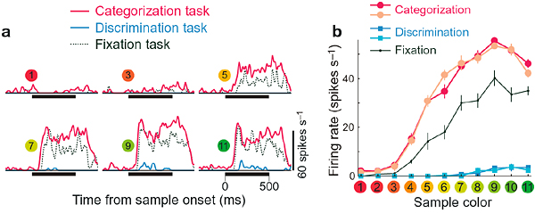 Two different functions can be distinguished in our color vision; one is categorization, the other is fine discrimination. These functions are utilized depending on the task demand, and we found that activities of many neurons in the inferior temporal cortex of the monkey to the same color stimulus changes depending on the task demands. Many neurons showed stronger responses when the monkey makes categorical color judgement than when it makes fine discrimination. This figure shows the responses of one such example of neuron. Responses during categorization, discrimination and fixation tasks are shown as histograms in a and as line graphs in b.
Staff
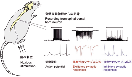
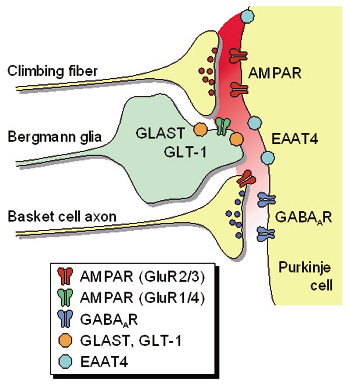
Our main interest lies in elucidation of the mechanism of transduction and integration of neural information in the nervous system. More specifically, we are trying to understand the basic properties of neural information processing between neurons or among a group of neurons constituting a local network. We are also interested in the pathophysiological mechanism how a single gene mutation leads to a symptom (such as ataxia and epilepsy), particularly in Ca2+ channel mutant mice. Additionally, we have recently started to make a computational approach, incorporating computer-based neurons into brain slice measurements (dynamic clamp), together with computational simulation of network functions. The following are currently ongoing major projects. (1) Studies of neurological disorders caused by calcium channel mutations. Mutations of the voltage-gated calcium channels are associated with neurological disorders of human and mice, which include cerebellar ataxia and some forms of seizure disorders. We study the relation how a single mutation causes neurological manifestations, mainly using brain slice preparations. (2) In vivo spinal nociceptive transmission and its plastic change. The spinal dorsal horn, especially superficial dorsal horn, has an important role in the transmission and modulation of nociceptive information. Superficial dorsal horn neurons make excitatory synaptic contacts with noxious primary afferent fibers. In addition, dorsal horn neurons receive inhibitory synaptic inputs from spinal interneurons and descending inhibitory neurons in the brain stem. We investigate the spinal modulatory mechanism of nociceptive transmission by using in vivo patch-clamp recording techniques (Fig. 2). We also study the underlying mechanism for the development of chronic pain. (3) Transmitter diffusion-mediated crosstalk between heterologous neurons. We previously reported that the excitatory transmitter glutamate diffused from climbing fiber (CF) terminals [projection to cerebellar Purkinje cells (PCs) from the inferior olive in the brain stem] presynaptically suppressed the inhibitory information flow from basket cells (BCs) to the same PCs. The heterosynaptic inhibition is mediated by the activation of presynaptic AMPA receptors expressed at the BC terminals. Recently, we found that glutamate transporters (EAAT4/GLAST/GLT-1) take unique part in determining the degree of CF-induced inhibition by influencing the glutamate concentration in the route of its extrasynaptic diffusion (Fig. 3).
Staff
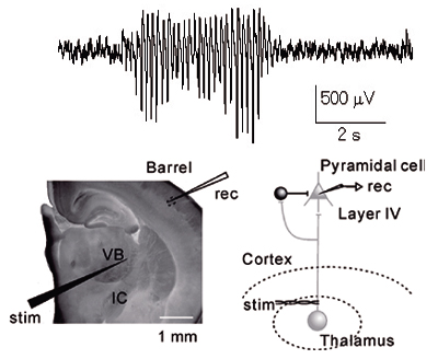
1. Analysis of the neuronal circuits in visual cortex Primary visual cortex is one of the best areas to study the relationship between brain functions and synapses/neural circuits, because the visual responsiveness of each neuron and the functional columnar structures are well characterized in this area. Therefore, we have analyzed the synapses and neuronal circuits in this cortical area, and clarified their basic properties. For example, we tested for fine-scale specificity of connections in rat visual cortex using cross-correlation analyses of synaptic currents evoked by laser scanning photostimulation. Recording simultaneously from adjacent layer 2/3 pyramidal cells, we found that when the cells were connected they shared inputs from individual excitatory neurons in layer 4 and layer 2/3. Thus, excitatory connections from layer 4 to layer 2/3 and within layer 2/3 form fine-scale assemblies of selectively interconnected neurons. To characterize connection properties further and elucidate the role of the fine-scale circuit in visual information processing, we are currently conducting electrophysiological analyses of neural circuits using slice preparations prepared from transgenic mice in which cells responding to particular visual stimulation can be visualized by fluorescent proteins expressed in an activity-dependent manner. 2. The activity-dependent developmental of visual responsiveness and neuronal circuits It is known that visual function matures under the strong influence of postnatal experience. We have been examining the effect of manipulation of visual inputs on the development of synaptic connections and neuronal circuits, to unravel the synaptic mechanisms of activity-dependent maturation of cortical functions. 3. Neuronal basis of locomotion and its development Recent molecular genetic studies suggest that the expression of transcription factors in the developing spinal cord helps determine the morphological and physiological properties of neurons. Using the zebrafish preparation, we have been examining the electrophysiological and morphological properties of neurons specified by individual or sets of transcription factors. 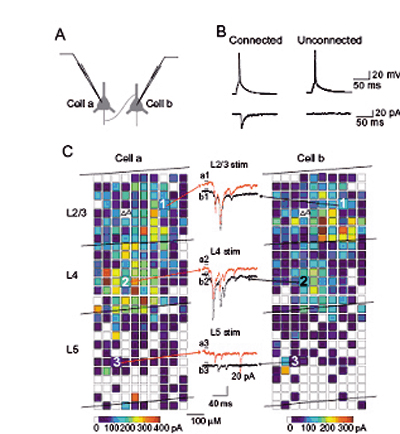 Analyses of photostimulation-evoked excitatory postsynaptic currents (EPSCs) simultaneously recorded in a pair of layer 2/3 pyramidal neurons that was synaptically connected. For each of the two cells, reconstructions of the locations of photostimulation sites (coloured squares) relative to the locations of laminar borders and cell bodies of recorded pyramidal neurons (triangles) are shown. The colour of each square indicates the sum of amplitudes of EPSCs that were observed in response to photostimulation at that site. 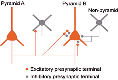 Inhibition between nearby pyramidal neurons via inhibitory synaptic terminals 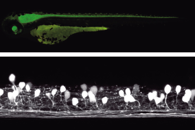 Studies with zebrafish as a model system to understand molecular mechanisms underlying development of neuronal wiring and neurophysiology of locomotion. In the transgenic zebrafish, a class of inteneurons are easily identified by fluorescence of GFP in live animals. The upper panel is an image using a regular fluorescent micoscope. The bottom panel is an image by a confocal microscopy. Staff
| ||||||||||||||||||||||||||||||||||||||||||||||