|
|
|
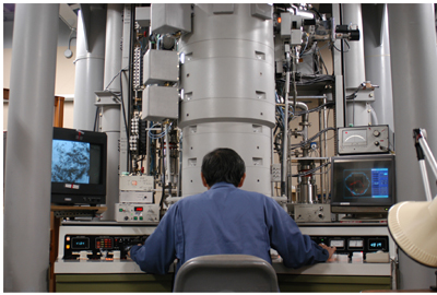
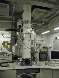
10. LARGE FACILITIES AND EQUIPMENTS FOR COOPERATIVE STUDIESAs a mission of NIPS to be the inter-university research institute, which conduct joint studies with researchers from domestic or foreign universities and other research institutes. NIPS provides specialized equipments, large-scale equipments and research facilities, and develops new equipments for morphological and functional 4D imagings of various organs such as brain. High voltage electron microscope (H-1250M; 1,000kV)
The high voltage electron microscope (H-1250M) has been specially designed for biological and medical research. The daily accelerating voltage of the microscope is 1,000kV. The pressure near the specimen position is less than 7×10⁻⁶ Pa and the magnification ranges from 1k to 1,000k times. Transmission images of thick specimens till about 5mm between ±60º tilt can be taken by side entry goniometer stage.
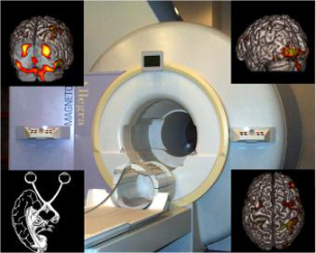
Magnetic Resonance Imaging System
MRI is an imaging technique that utilizes the nuclear magnetic resonance of the hydrogen atom. Not only to image the anatomical details of the brain, MRI allows to explore the neural substrates of human cognitive function by the visualization of the task-related changes in regional cerebral blood flow (functional MRI). NIPS installed 3 Tesla magnetic resonance imaging system (Allegra, Siemens) in FY 2000.
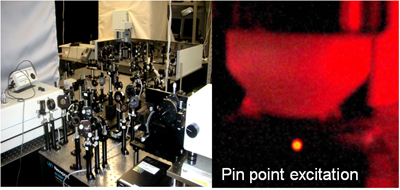
Mutiphoton excitation microscopy Multiphoton excitation of photo-sensitive molecules e.g. fluorescent molecules, at a limited area with a high density of photon is introduced by a high power femtosecond pulse laser. Miltiphoton microscopy allows us to observe fine structures and their mobility, e.g. synaptic structures and various cells, as well as neuronal activity, in an in vivo and in vitro preparations.
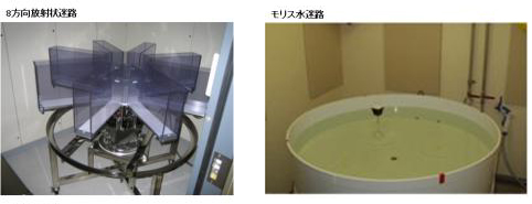
A comprehensive behavioral test battery In our laboratory, we can conduct various kinds of behavioral tests for genetically engineered mice, including wire hang, grip strength, light/dark transition, open field, elevated plus maze, hot plate, social interaction, rotarod, prepulse inhibition/startle response, Porsolt forced swim, gait analysis, eight-arm radial maze, T maze, Morris water maze, Barnes maze, object recognition test, cued and contextual fear conditioning, passive avoidance, tail suspension, and 24 hour home cage monitoring .
The primary goal of our research group is to reveal functional significances of genes and their involvement in neuropsychiatric disorders by conducting a comprehensive behavioral test battery on genetically engineered mice. |
|
|
|
| Copyright(C) 2009 NIPS (National Institute for Physiological Sciences) | |