10. LARGE FACILITIES AND EQUIPMENTS FOR COOPERATIVE STUDIES
As a mission of NIPS to be the inter-university research institute, which conduct joint studies with researchers from domestic or foreign universities and other research institutes. NIPS provides specialized equipments, large-scale equipments and research facilities, and develops new equipments for morphological and functional 4D imagings of various organs such as brain.
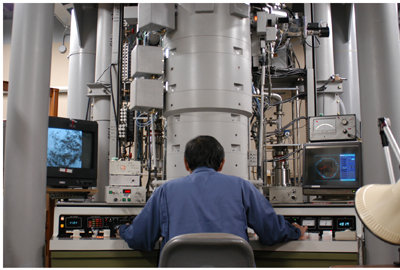
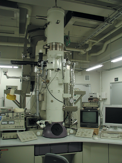
High voltage electron microscope (HVEM)
The high voltage electron microscope (H-1250M) is specially designed for biological and medical research. The daily accelerating voltage of the microscope is 1,000kV. The pressure of less than 7×10⁻⁶ Pa near the specimen position, performs the observation of biological samples at the magnification ranges from 1k to 1,000k times. Transmission images of thick specimens till about 5mm are collected between ±60º tilt by the side entry goniometer stage.
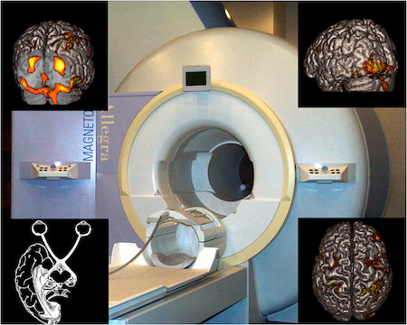
Magnetic Resonance Imaging System
MRI is an imaging technique that utilizes the nuclear magnetic resonance of the hydrogen atom. Not only to image the anatomical details of the brain, MRI allows to explore the neural substrates of human cognitive function by the visualization of the task-related changes in regional cerebral blood flow (functional MRI).
NIPS installed 3 Tesla magnetic resonance imaging system (Allegra, Siemens) in FY 2000.
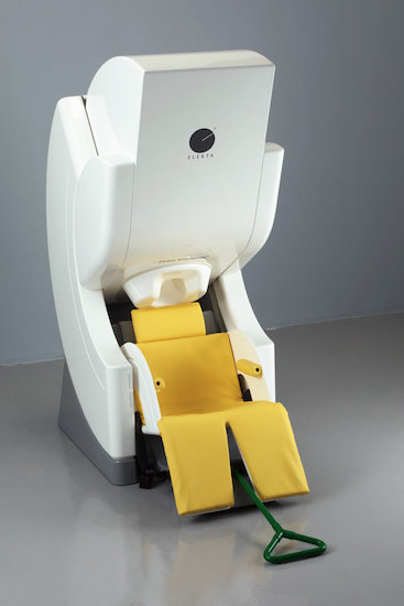
Magnetoencephalography (MEG)
Magnetoencephalography (MEG) has potential to measure brain activities with better temporal and spatial resolution in milliseconds and millimeter, respectively, compared with other methods such as functional magnetic resonance imaging. Event-related magnetic fields following various kinds of sensory stimulation are mainly analyzed. In addition, background brain activities (brain waves) in various conditions can be analyzed.
Phase Contrast Cryo-electron Microscope
This is an electron microscope developed for observing a close-to-life state of biological samples without harsh sample treatments such as dehydration, plastic embedding and staining. Biological sample specimens with a thickness up to 500nm can be observed with a high contrast without staining. Structural analyses for protein molecules, viruses, bacteria, cultured cells and tissue sections are the target of this novel microscopic system.
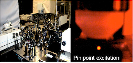
Mutiphoton excitation microscopy
Multiphoton excitation of photo-sensitive molecules e.g. fluorescent molecules, at a limited area with a high density of photon is introduced by a high power femtosecond pulse laser. Miltiphoton microscopy allows us to observe fine structures and their mobility, e.g. synaptic structures and various cells, as well as neuronal activity, in an in vivo and in vitro preparations.
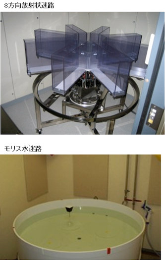
A comprehensive behavioral test battery
In our laboratory, we can conduct various kinds of behavioral tests for genetically engineered mice, including wire hang, grip strength, light/dark transition, open field, elevated plus maze, hot plate, social interaction, rotarod, prepulse inhibition/startle response, Porsolt forced swim, gait analysis, eight-arm radial maze, T maze, Morris water maze, Barnes maze, object recognition test, cued and contextual fear conditioning, passive avoidance, tail suspension, and 24 hour home cage monitoring.
The primary goal of our research group is to reveal functional significances of genes and their involvement in neuropsychiatric disorders by conducting a comprehensive behavioral test battery on genetically engineered mice.
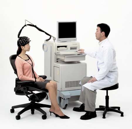
NIRS (Near infrared spectroscopy)
This is one of hemodynamic methods to investigate human brain function using near infrared spectroscopy (NIRS). Advantages of NIRS are as follows. (1) This is a completely non-invasive method. (2) This method is available for subjects who are not restricted, being completely different from fMRI, PET and MRG. Therefore, for example, brain activity during walking and cooking can be analyzed. (3) NIRS can be recorded for infants, since subjects do not have to be restricted. NIPS has recording probes for both adults and infants.