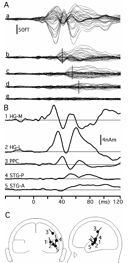
Although anatomical, histochemical and electrophysiological findings in both animals and humans have suggested a parallel and serial mode of auditory processing, precise activation timings of each cortical area are not well known, especially in humans. We investigated the timing of arrival of signals to multiple cortical areas using magnetoencephalography in humans. Following click stimuli applied to the left ear, activations were found in six cortical areas in the right hemisphere: the posteromedial part of Heschl's gyrus (HG) corresponding to the primary auditory cortex (PAC), the anterolateral part of the HG region on or posterior to the transverse sulcus, the posterior parietal cortex (PPC), posterior and anterior parts of the superior temporal gyrus (STG), and the planum temporale (PT). The mean onset latencies of each cortical activity were 17.1, 21.2, 25.3, 26.2, 30.9 and 47.6 ms respectively. These results suggested a serial model of auditory processing along the medio-lateral axis of the supratemporal plane and, in addition, implied the existence of several parallel streams running postero-superiorly (from the PAC to the belt region and then to the posterior STG, PPC or PT) and anteriorly (PAC-belt-anterior STG).
Inui K, Okamoto H, Miki K, Gunji A, Kakigi R: Serial and parallel processing in the human auditory cortex: a magnetoencephalographic study. Cerebral Cortex. Jan;16(1):18-30

Multi-dipole analysis of magnetic fields later than 1M. A: waveforms of recorded magnetic fields (a) and residual magnetic fields obtained by subtraction of theoretical magnetic fields due to the model in each step from the recorded magnetic fields (b-e). B: time course of each source activity. C: schematic drawings of the location and orientation of each source. HG, Heschl's gyrus; PPC, posterior parietal cortex; STG, superior temporal gyrus.

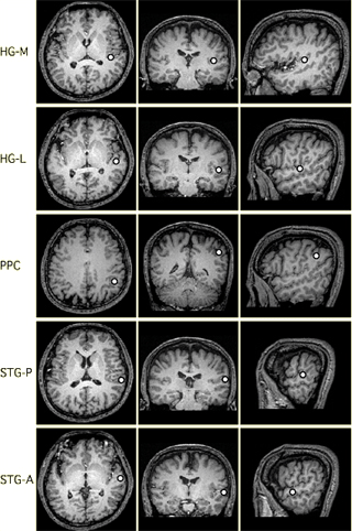
Locations of each cortical source in Figure 1 superimposed on the subject's MR images.