The go ⁄ nogo task is a useful paradigm for recording event-related potentials (ERPs) to investigate the neural mechanisms of response inhibition. In nogo trials, a negative deflection at around 140-300 ms (N2), which has been called the 'nogo potential', is elicited at the frontocentral electrodes, compared with ERPs recorded in go trials. In the present study, we investigated the generators of nogo potentials by recording ERPs and by using magnetoencephalography (MEG) simultaneously during somatosensory go ⁄ nogo tasks to elucidate the regions involved in generating nogo potentials. ERP data revealed that the amplitude of the nogo-N140 component, which peaked at about 155 ms from frontocentral electrodes, was significantly more negative than that of go-N140. MEG data revealed that a long-latency response peaking at approximately 160 ms, termed nogo-M140 and corresponding to nogo-N140, was recorded in only nogo trials. The equivalent current dipole of nogo-M140 was estimated to lie around the posterior part of the inferior frontal sulci in the prefrontal cortex. These results revealed that both nogo-N140 and nogo-M140 evoked by somatosensory go ⁄ nogo tasks were related to the neural activity generated from the prefrontal cortex. Our findings combining MEG and ERPs clarified the spatial and temporal processing related to somato-motor inhibition caused in the posterior part of the inferior frontal sulci in the prefrontal cortex in humans.
>Nakata H, Inui K, Wasaka T, Akatsuka K & Kakigi R Somato-motor inhibitory processing in humans: A study with MEG and ERP. Eur J Neurosci, 22(7): 1784-1792
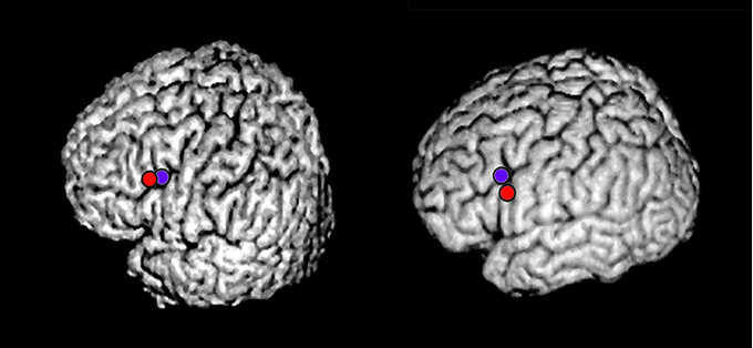
Nogo-dominant activity in a three-dimensional image of a representative subject. The activity was estimated around the posterior part of the inferior frontal sulci in the left prefrontal cortex. Red and blue points indicate the activities for the left second and fifth digits, respectively.


