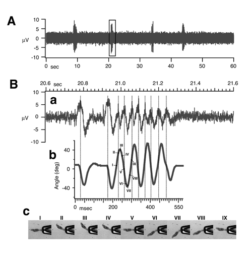
To examine the role of the amino acid GABA in the locomotion of basal chordates, we investigated the pharmacology of swimming and the morphology of GABA-immunopositive neurones in tadpole larvae of the ascidians Ciona intestinalis and Ciona savignyi. We verified that electrical recording from the tail reflects alternating muscle activity during swimming by correlating electrical signals with tail beats using high-speed video recording. GABA reversibly reduced swimming periods to single tail twitches, while picrotoxin increased the frequency and duration of electrical activity associated with spontaneous swimming periods. Immunocytochemistry for GABA revealed extensive labelling throughout the larval central nervous system. Two strongly labelled regions on either side of the sensory vesicle were connected by an arc of labelled fibres, from which fibre tracts extended caudally into the visceral ganglion. Fibre tracts extended ventrally from a third, more medial region in the posterior sensory vesicle. Two rows of immunoreactive cell bodies in the visceral ganglion extended neurites into the nerve cord, where varicosities were seen. Thus, presumed GABAergic neurones form a network that could release GABA during swimming that is involved in modulating the time course and frequency of periods of spontaneous swimming. GABAergic and motor neurones in the visceral ganglion could interact at the level of their cell bodies and/or through the presumed GABAergic fibres that enter the nerve cord. The larval swimming network appears to possess some of the properties of spinal networks in vertebrates, while at the same time possibly showing a type of peripheral innervation resembling that in some protostomes.
Brown, E.R., Nishino, A., Bone, Q., Meinertzhagen, I.A., Okamura, Y. Eur J Neurosci 22, 2541-2548 (2005)

Muscle field potentials recorded from the Ciona intestinalis larval tail report its contractile states. (A) Example of muscle field potentials recorded for 1 min. High-speed video images were simultaneously captured during the second period of swimming (rectangle). (B) (a) Expanded trace of the second period of field potentials shown in A. (b) Angles of tail inflections, plotted as the subtense of the line from the tip of the larva to the axis of the glass pipette are plotted at 2.5 msec intervals. Each cycle of inflection corresponds well to field potential activity (dotted lines). (c) Images of the larva during a single cycle of inflection (I-IX, indicated in b) are shown at 10 msec intervals.