Shimada A, Niwa H, Tsujita K, Suetsugu S, Nitta K, Hanawa-Suetsugu K, Akasaka R, Nishino Y, Toyama M, Chen L, Liu ZJ, Wang BC, Yamamoto M, Terada T, Miyazawa A, Tanaka A, Sugano S, Shirouzu M, Nagayama K, Takenawa T, and Yokoyama S, (2007) “Curved EFC/F-BAR-Domain Dimers Are Joined End to End into a Filament for Membrane Invagination in Endocytosis”, Cell 129, 761-772.
RIKEN press-released a breakthrough on one of the basic cellular processes, endocytosis, which was achieved as a collaboration of the other two institutions and published in the recent issue of Cell. In this research topics the particular contribution of Nano-Structure Physiology Lab to the achievement is focused.
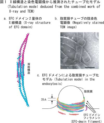
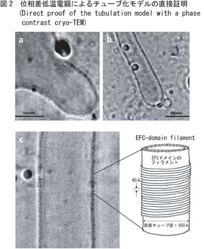
Endocytosis is a complicated process where first the cell surface sinks, second fine particles like pathogenic germs such as viruses are wrapped by surface membrane, third fine particles by membrane is hollowed in a tubular form and finally membrane-wrapped particles are teared off. In the series of the endocytotic process, the mechanism underlying the third step has long been uncovered. Yokoyama group of RIKEN has been studying the key protein "EFC domain" assumed to have a function of the membrane tabulation. Recently its atomic structure was solved by X-ray crystallography and the elongated bow-shape was known (Fig. 1a). On the other hand conventional transmission electron microscopy (TEM) with staining has shown a liposomal tabulation induced by the key protein (Fig. 1b). Combining two results, a model for membrane tabulation was proposed (Fig. 1c). But its experimental verification has not yet made due to the limitation of conventional TEM. The pitfall has been avoided by an application of phase contrast cryo-TEM, which enables to observe specimens at a nanometer resolution in their intact form without staining. The results are shown in Fig. 2 as the initial process of the tabulation (Figs. 2a, b) and high resolution details (Fig. 2c) confirming the predicted model (Fig. 1c).
This achievement will give a clue to the medical care for virus or bacterial infectious diseases.