Dutta AK, Korchev YE, Shevchuk AI, Hayashi S, Okada Y, Sabirov RZ. Spatial distribution of maxi-anion channel on cardiomyocytes detected by smart-patch technique.
Biophys J. 2008 [Published ahead of print on November 16, 2007 as doi:10.1529/biophysj.107.117820]
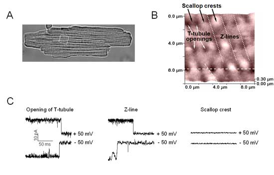
We have recently determined that the maxi-anion channel is a nanoscopic pore well suited to function as an ATP-releasing pathway. Studying cardiac cells, we have demonstrated that the ATP release from neonatal rat cardiomyocytes is mainly mediated by activity of the maxi-anion channel under ischemic or hypotonic conditions. Paradoxically, these channels could not be observed in cardiomyocytes freshly isolated from adult hearts with commonly used patch-clamp conditions. It was therefore suggested that maxi-anion channels are only transiently expressed in neonatal cells, disappearing upon maturation. However, ATP release from mature cardiomyocytes and purinergic signaling in the normal and diseased heart are well-recognized phenomena. We thus hypothesized that the difference in maxi-anion channel activity between neonatal and adult cells could be related to different pattern of spatial distribution of the maxi-anion channels over the surface of sarcolemma. In the present study, the spatial distribution of maxi-anion channels and that of ATP release site were first studied in neonatal rat cardiomyocytes by using a recently developed “smart-patch” method and an ATP biosensor technique, respectively. Both distributions matched with each other providing further evidence for the “maxi-anion channel = ATP-conductive channel” concept. We then reexamined functional expression of maxi-anion channels in adult rat cardiomyocytes by the “smart-patch” technique using very fine-tipped patch pipettes. When fine-tipped 15-20 MΩ pipettes were targeted to only Z-line areas, we observed, for the first time, the maxi-anion events. Smart-patching different regions of the cell surface, we found that the channel activity only at the openings of T-tubules and along Z-lines but not in the scallop crest area. Thus, it is concluded that maxi-anion channels are concentrated at the openings of T-tubules and along Z-lines in adult cardiomyocytes. Our study showed that the “smart-patch” technique provides a powerful method to detect a unitary event of channels which are localized at some specific site in the narrow region.