Endo T1,4, Tarusawa E2, Notomi T2, Kaneda K1,4, Hirabayashi M, S3higemoto R2, Isa T1,4 (2008) Dendritic Ih Ensures High-fidelity Dendritic Spike Responses of Motion Sensitive Neurons in Rat Superior Colliculus. Journal of Neurophysiology, Jan 23; [Epub ahead of print]
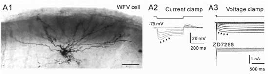
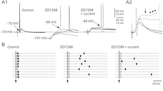
Hyperpolarization-activated cyclic nucleotide-gated (HCN) channels that generate Ih currents are widely distributed in the brain, and have been shown to contribute to various neuronal functions. In the present study, we investigated the functions of Ih in the motion sensitive projection neurons (WFV cells) of the superior colliculus, a pivotal visual center for detection of and orientating to salient objects. Combination of whole-cell recordings and immunohistochemical investigations suggested that WFV cells express HCN1 channels mainly on their expansive dendritic trees. We found that blocking Ih suppressed the initiation of short- and fixed-latency dendritic spike responses, and led instead to long- and fluctuating-latency somatic spike responses to optic fiber stimulations. These results suggest that the dendritic Ih facilitates the dendritic initiation and⁄or propagation of action potentials and ensures that WFV cells generate spike responses to distal synaptic inputs in a sensitive and robustly time-locked manner, probably by acting as continuous depolarizing drive and fixing dendritic membrane potentials close to the spike threshold. These functions are different from known functions of dendritic Ih revealed in hippocampal and neocortical pyramidal cells, where they spatiotemporally limit the propagations of synaptic inputs along the apical dendrites by reducing dendritic membrane resistance. Thus, we have revealed new functional aspects of Ih, and these dendritic properties are likely critical for visual motion processing in these neurons.