During a visual search, information about the visual attributes of objects and behavioral requirements is essential for discriminating a target object from others in the visual field. On the other hand, information about the object’s position appears to be more important when orienting the eyes toward the target. To understand the neural mechanisms underlying such a transition (i.e., from nonspatial, feature-based target selection to spatial-based target selection), we examined the dependence of neuronal activity in the macaque posterior parietal cortex (PPC) on visual sensory properties and ongoing task demands. Monkeys were trained to perform a visual search task in which either a shape or color singleton within an array was the target, depending on the ongoing search dimension. The visual properties and the task demands were manipulated by independently changing the stimulus features (shape and color), singleton type, and search dimension. We found that a subset of PPC neurons significantly discriminated the target from other stimuli only when the target was defined by a particular stimulus dimension and had specific stimulus features, such as a shape-singleton, bar stimulus (conditiondependent target selection), whereas another subset did so irrespective of the stimulus features and the target-defining dimension (conditionindependent target selection). There was thus a great deal of variety in the neural representations specifying the locus of the target. The coexistence of these distinctly different types of target-discrimination processes suggests the PPC may be situated at the level where the transition from nonspatial- to spatial-based target selection takes place.
Ogawa T and Komatsu H
Condition-dependent and condition-independent target selection in the macaque posterior parietal cortex.
J Neurophysiology, 101:721-736, 2009.
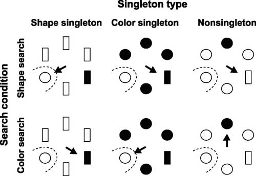
Experimental paradigm and visual stimuli. Stimulus conditions for the experiment. Two singleton stimuli, one unique in the color dimension, the other in the shape dimension, were presented simultaneously with four additional identical stimuli. Open and filled symbols correspond to cyan and yellow, respectively. Monkeys had to make a saccade (arrows) to one of the singleton stimuli, depending on the instructed target-defining dimension. In the shape search (top row), the shape singleton was the target and the color singleton was the distractor. In the color search, the shape singleton was the distractor and the color singleton was the target. The nonsingleton stimulus never became the target (nontarget). Examination of the 2 search conditions was conducted in separate blocks. The ongoing target-defining dimension was signaled by the color of the fixation spot.
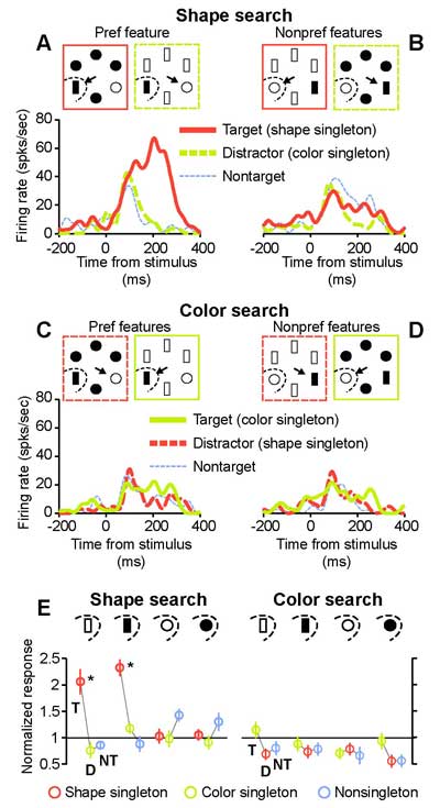
A parietal neuron showing dependence on stimulus features and the target-defining dimension. A-D: spike density functions during the multidimensional visual search task. Thick red and green lines indicate the responses to the shape and color singleton stimuli appearing in the receptive field. Dotted thin blue lines indicate the responses to a nontarget (nonsingleton) stimulus. Solid and dashed lines, respectively, indicate that the stimulus in the receptive field was the target or distractor. The responses are temporally aligned at the onset of the stimulus array. The spike density functions were smoothed with a Gaussian function (SD = 10 ms). Top insets in each panel show the stimulus conditions under which the stimulus in the receptive field was the target or distractor. Formats of the rectangular frames in the insets are the same as in the spike density functions. This neuron only exhibited a marked response to the target when a specific combination of the stimulus feature (bar shape) and the target-defining dimension (shape dimension) was met (A). E: normalized mean responses (with SE) of the same neuron for the entire set of 24 trial conditions, which were calculated by dividing the responses to the 24 different stimulus conditions by their average. Top insets indicate the stimulus features in the receptive field in the shape search (left) and the color search (right). Data for the same stimulus features are connected by lines under each search condition. In each set, 3 connected circles respectively indicate from left to right that the stimulus in the receptive field was the target (T), distractor (D), or nontarget (NT). Red, green, and blue circles represent the responses to the shape singleton, the color singleton, and the nonsingleton stimuli, respectively. Asterisks indicate significant difference from the 2 other responses in each set (Mann-Whitney U test, P < 0.01).
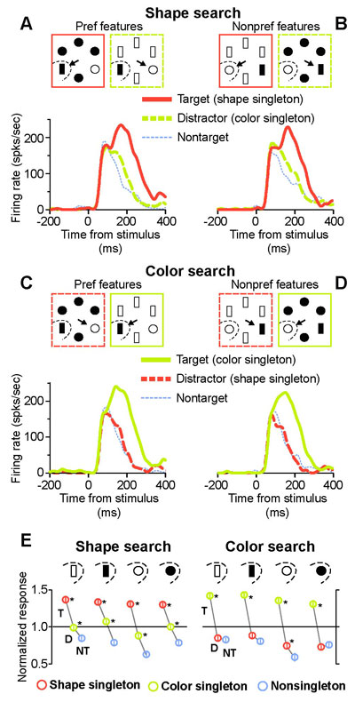
A parietal neuron showing no dependence on stimulus features or the target-defining dimension. The activity of this neuron always exhibited significant discrimination of the target from the other stimuli, irrespective of the stimulus features and target-defining dimension. Conventions are the same as in Fig. 2.
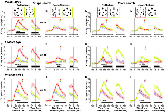
Time course of the population responses. Average population responses of the variant-type neurons (n = 21) in the shape search (A, B) and the color search (C, D). For clarity, only neurons that preferred the shape dimension are shown. The stimulus shown in the receptive field had either the preferred (A, C) or nonpreferred (B, D) stimulus features. Traces in each panel are aligned at the time of array presentation (left) or saccade initiation (right). Thick solid, thick dashed, and thin dotted lines indicate the population responses to the target, distractor, and nontarget stimuli, respectively. Red, green, and blue lines respectively correspond to the responses to a shape, color, and nonsingleton stimuli. Shaded areas indicate 1SE. Red, green, and blue triangles respectively denote mean saccade reaction times when a shape, color, or nonsingleton stimulus was presented within the receptive field. Horizontal black lines below the spike density functions indicate the period during which the target was significantly discriminated from the distractor and nontarget stimuli (permutation test, P < 0.01). The number above this horizontal line indicates the first time after the stimulus onset that target discrimination became significant and continued for > 50 ms. E-H: time courses of the population responses of the feature-type neurons (n = 11) are shown in the same format as in A-D. I-L: time course of the population responses of the invariant-type neurons (n = 35).


