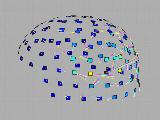
MEG response waveform carries direction information of visually presented motion stimulus. This figure shows 3-dimensional illustration of the current vectors (shown by the colored lines) for 4 motion directions measured at each sensor location shown by the square at around the peak of the response (viewing from left posterior). The color of the line indicates motion direction (cyan: rightward; magenta: upward; white: leftward; yellow: downward) and the length corresponds to the current moment. The color of the square shows the magnetic field strength (that increases from blue to red) measured at the sensor.
Kaneoke Y, Urakawa T, and Kakigi R.
Visual motion direction is represented in population-level neural response as measured by magnetoencephalography.
Neuroscience 160, 676-687, 2009.
We investigated whether direction information is represented in the population-level neural response evoked by the visual motion stimulus, as measured by magnetoencephalography. Coherent motions with varied speed, varied direction, and different coherence level were presented using random dot kinematography. Peak latency of responses to motion onset was inversely related to speed in all directions, as previously reported, but no significant effect of direction on latency changes was identified. Mutual information entropy (IE) calculated using four-direction response data increased significantly (>2.14) after motion onset in 41.3% of response data and maximum IE was distributed at approximately 20 ms after peak response latency. When response waveforms showing significant differences (by multivariate discriminant analysis) in distribution of the three waveform parameters (peak amplitude, peak latency, and 75% waveform width) with stimulus directions were analyzed, 87 waveform stimulus directions (80.6%) were correctly estimated using these parameters. Correct estimation rate was unaffected by stimulus speed, but was affected by coherence level, even though both speed and coherence affected response amplitude similarly. Our results indicate that speed and direction of stimulus motion are represented in the distinct properties of a response waveform, suggesting that the human brain processes speed and direction separately, at least in part.


