To examine the intrasynaptic arrangement of postsynaptic receptors in relation to the functional role of the synapse, we quantitatively analyzed the two-dimensional distribution of AMPA and NMDA receptors (AMPARs and NMDARs, respectively) using SDS-digested freeze-fracture replica labeling (SDS-FRL) and assessed the implication of distribution differences on the postsynaptic responses by simulation. In the dorsal lateral geniculate nucleus, corticogeniculate (CG) synapses were twice as large as retinogeniculate (RG) synapses but expressed similar numbers of AMPARs. Two-dimensional views of replicas revealed that AMPARs form microclusters in both synapses to a similar extent, resulting in larger AMPAR-lacking areas in the CG synapses. Despite the broad difference in the AMPAR distribution within a synapse, our simulations based on the actual receptor distributions suggested that the AMPAR quantal response at individual RG synapses is only slightly larger in amplitude, less variable, and faster in kinetics than that at CG synapses having a similar number of the receptors. NMDARs at the CG synapses were expressed twice as many as those in the RG synapses. Electrophysiological recordings confirmed a larger contribution of NMDAR relative to AMPAR-mediated responses in CG synapses. We conclude that synapse size and the density and distribution of receptors have minor influences on quantal responses and that the number of receptors acts as a predominant postsynaptic determinant of the synaptic strength mediated by both the AMPARs and NMDARs.
Etsuko Tarusawa, Ko Matsui*, Timotheus Budisantoso, Elek Molnár, Masahiko Watanabe, Minoru Matsui, Yugo Fukazawa*, Ryuichi Shigemoto (2009)
Input-Specific Intrasynaptic Arrangements of Ionotropic Glutamate Receptors and Their Impact on Postsynaptic Responses.
The Journal of Neuroscience, 29(41):12896-12908.
(* Corresponding authors)
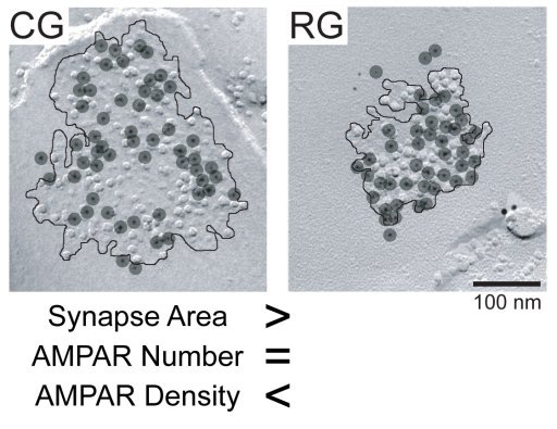
SDS-digested freeze-fracture replica labeling (SDS-FRL) images of synapses on relay cells in the dorsal lateral geniculate nucleus (dLGN). Examples from a corticogeniculate (CG) synapse and a retinogeniculate (RG) synapse are shown. Synaptic area is demarcated by a black line and the location of the AMPA-type glutamate receptor (AMPAR) labeling is shown with black dots. CG synapse had larger area, however, the number of AMPAR labeling was similar between the two synapses, thereby resulting in a higher AMPAR density in the RG synapse. In addition, areas devoid of apparent AMPAR labeling were often found in the CG synapses suggesting that AMPAR distribution is non-uniform.
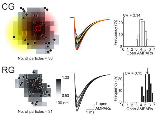
Glutamate diffusion and receptor response was simulated based on the AMPAR distribution found in CG and RG synapses using SDS-FRL. AMPAR locations are shown as black dots on the left column figure. Synaptic response caused by glutamate released in vicinity to an AMPAR receptor cluster (red square) and in an open space devoid of AMPARs (yellow square) are shown as the red trace and the yellow trace, respectively, in the middle column figure. The amplitude of the synaptic response to releases at various locations within the synaptic contact is expressed in gray scale on the left column figure. The right column figure is the amplitude histogram. Based on these simulations, we found that 1) the synaptic response depends only slightly on the release location, 2) although CG and RG synapses had difference in the AMPAR density, the synaptic responses were nearly the same, 3) the major factor determining the size of the synaptic response is the number of receptors expressed.


