Localization of NK1 receptors and roles of substance-P in subepithelial fibroblasts of rat intestinal villi.
Furuya S, Furuya K, Shigemoto R, Sokabe M.
Cell Tissue Res (2010) 342:243-259
Subepithelial fibroblasts of the intestinal villi, which form a contractile cellular network beneath the epithelium, are in close contact with epithelial cells, nerve varicosities, capillaries, smooth muscles and immune cells, and secrete extracellular matrix molecules, growth factors and cytokines, etc. Cultured subepithelial fibroblasts of the rat duodenal villi display various receptors such as endothelins, ATP, substance-P
and bradykinin, and release ATP in response to mechanical stimulation. In this study, the presence of NK1 receptors (NK1R) in the villous subepithelial fibroblasts was confirmed by Ca2+ measurement and immunohistochemistry. These villous subepithelial fibroblasts form synapse-like structures with both substance-P –immunopositive (afferent neuron) and -immunonegative nerve varicosities. The mutual interaction between subepithelial fibroblasts and afferent neurons via ATP and substance-P plays a key role in the structure and function of the intestinal villi.
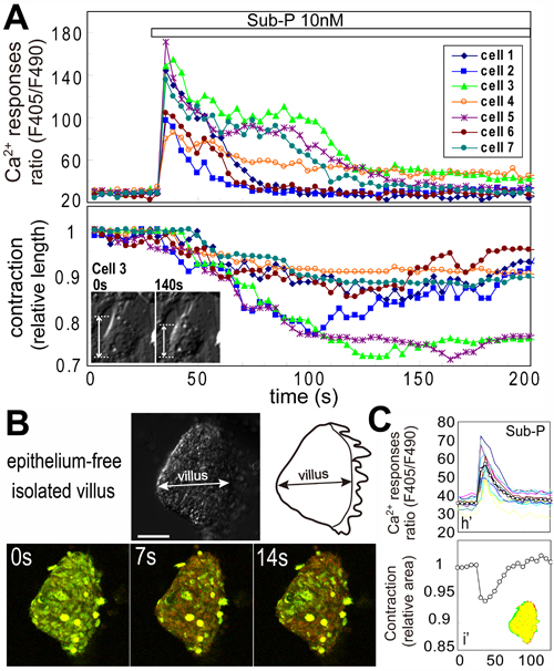
Substance-P elicited an increase in the intracellular Ca2+ concentration followed contraction of the subepithelial fibroblasts in primary culture isolated from rat duodenum (A). Newly developed preparations of epithelium-free isolated villi (B) respond to substance-P and the Ca2+ increase and the contraction of villi were observed (B, C).
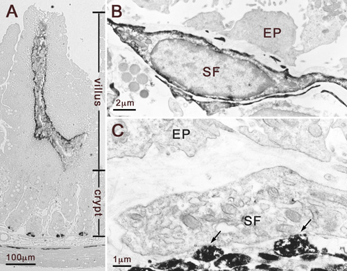
The localization of NK1R and substance-P in the villi was examined by light (A) and electron (B) microscopic immunohistochemistry. NK1R immunoproducts (dark DAB products) were intensely localized on the plasma membrane of villous subepithelial fibroblasts (A, B), but pericryptal fibroblasts were NK1R immuno-negative (A). Villous subepithelial fibroblasts form synapse-like structure (arrows) with substance-P immunopositive nerve terminals (marked by dark DAB products)