Retinal bipolar, amacrine, and ganglion cells contact each other within precisely defined synaptic laminae, but the spatial distribution of contacts between the cells is generally treated as random. Here we show that not to be the case. Excitatory inputs to inner retinal neurons were visualized by introduction of a plasmid coding for the postsynaptic protein PSD95-GFP. Our initial finding was that synapses on the dendrites of retinal ganglion cells are regularly spaced, at 2-3-μm intervals, along the dendrites. Thus, the presence of a PSD95 punctum creates a nearby zone from which other inputs appear to be excluded. Despite their great variation in size and different morphologies, the spacing is similar for the arbors of different retinal ganglion cell types. Regular spacing was also observed for the starburst amacrine cells. This regularity is mirrored in the spacing of axonal varicosities of the stratified bipolar cells, which have a regular, nonrandom interval consistent with that of the PSD95 puncta on ganglion cells. Thus, for each level of the inner plexiform layer all three cell types participate in a single 2D mosaic of synaptic contacts. These findings raise a new set of questions: How does the self-avoidance of synaptic sites along an individual dendrite arise and how is it physically maintained? Why is a regular spacing of inputs important for the computational function of the cells? Finally, which of the three players, if any, is developmentally responsible for the initial establishment of the pattern?
J Comp Neurol. 2011 Feb 1;519(2):341-57.
Regular mosaic of synaptic contacts among three retinal neurons.
Koizumi A, Jakobs TC, Masland RH.
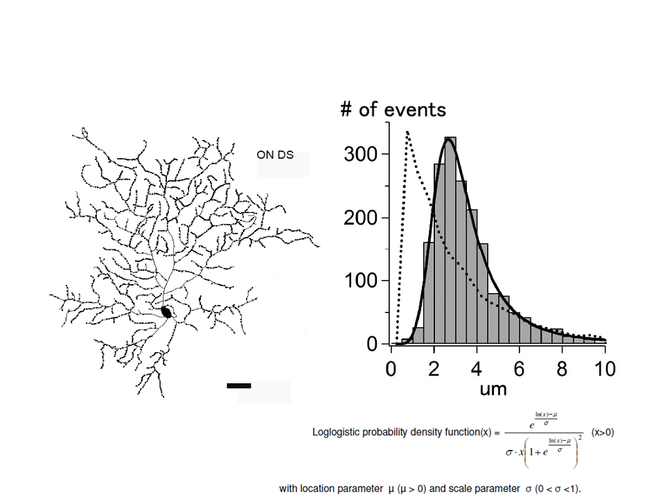
Figure 1. Excitatory synapses on dendrites of On-type DS ganglion cell and its one-dimensional nearest neighbor analysis.


