Damage to the cerebral cortex caused by stroke in adulthood often leads to impairment of contralateral limb movements. On the other hand, it has been reported that similar brain damage at the juvenile stage results in less severe impairment. It was shown that adult rodents with neonatal cortical damage exhibited fairly normal motor functions in their limbs on the contralateral side to the damage, while animals with a similar injury at maturity showed poor recovery. As a neuronal mechanism explaining this, corticospinal fibers from the sensorimotor cortex on the opposite side of the lesion issued collateral sprouting to the gray matter of the ipsilateral spinal cord, which could mediate cortical excitation to ipsilateral forelimb motoneurons (Takahashi et al. 2009, Umeda et al. 2010). However, it is not clear what cortical neurons issued aberrant axonal fibers to the ipsilateral spinal cord.
In this study, we identified origins of the ipsilateral or contralateral corticospinal projection by injecting retrograde neural tracers with different fluorescent probes into either side of the cervical spinal gray matter in neonatally hemidecorticated rats. We clarified that double-labeled neurons were rarely found in the undamaged cortex, which indicated that ipsilateral projections were not collateral of normal contralateral projections. Corticospinal neurons with contralateral or ipsilateral projections were distributed both in the rostral forelimb motor area (RFA) and the caudal forelimb motor area (CFA). While the distribution of the ipsilateral projection neurons largely overlapped that of the contralateral projection neurons in the RFA, the ipsilateral projection neurons tended to be segregated from the contralateral projection neurons in the CFA. The results suggested that the RFA and the CFA contribute to the compensatory process in different ways.
Tatsuya Umeda, Tadashi Isa
Differential contributions of rostral and caudal frontal forelimb areas to compensatory process after neonatal hemidecortication in rats.
Eur. J. Neurosci. 34 (2011) 1453-1460
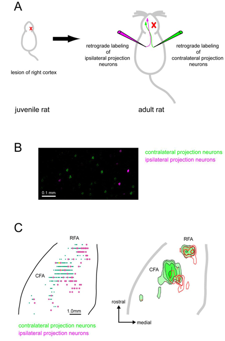
(A) Experimental design. Right cortex of the juvenile rat was lesioned on the 5th postnatal day. After maturation, the corticospinal neurons were retrogradely labeled using neuronal tracers with different fluorescent probes (green and magenta).
(B) Retrogradely labeled corticospinal neurons in the undamaged cortex of hemidecorticated rats. Contralateral projection neurons; green, ipsilateral projection neurons; magenta.
(C) Distributions of retrogradely labeled corticospinal neurons. Left; Two-dimensional schematic representations of the distributions of retrogradely labeled corticospinal neurons. Each dot represents single labeled neuron. Contralateral projection neurons; green, ipsilateral projection neurons; magenta. Right; Density maps of retrogradely labeled corticospinal neurons. Contralateral projection neurons; green, ipsilateral projection neurons; red.