Large facilities and equipments for cooperative studies
High voltage electron microscope (HVEM)
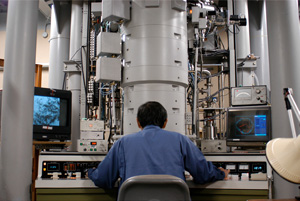
Hitachi H-1250M is the high voltage electron microscope specially designed for biological and medical sciences. The microscope usually operates at an accelerating voltage of 1,000kV. The column pressure is less than 7×10⁻⁶ Pa near the specimen position. Image acuisition of biological samples performs at the magnification ranges from 1k to 1,000k. Projections of thick biological specimens up to 5μm are collected at tilt angles between ±60° using the side entry specimen holder, which gives 3-dimentional ultra-structures of biological specimens at nano meter scales.
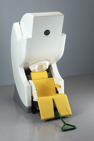
Magnetic Resonance Imaging System
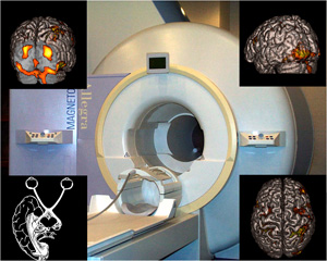
MRI is an imaging technique that utilizes the nuclear magnetic resonance of the hydrogen atom. Not only to image the anatomical details of the brain, MRI allows to explore the neural substrates of human cognitive function by the visualization of the task-related changes in regional cerebral blood flow (functional MRI).
For over a decade, we have been working on 3T MRI to investigate higher brain function of human. To simultaneously measure the neural activities of two participants during their social interaction,we have recently installed dual functional MRI system with two 3T MRI. Thus NIPS is now equipped with three 3-Tesla MRIs (Allegra, Siemens in FY 2000, and Verio x 2, Siemens in FY 2009).
Magnetoencephalography (MEG)
Magnetoencephalography (MEG) has potential to measure brain activities with better temporal and spatial resolution in milliseconds and millimeter, respectively, compared with other methods such as functional magnetic resonance imaging.Event-related magnetic fields following various kinds of sensory stimulation are mainly analyzed. In addition, background brain activities (brain waves) in various conditions can be analyzed.
Phase Contrast Cryo-electron Microscope
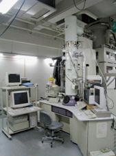
Phase contrast electron cryomicroscope is an electron microscope developed for observing close-to-life state biological samples with a combination of rapid freezing and ice embedding sample preparation methods. Biological specimens up to 200 nm thickness can be observed with a high-resolution and a high contrast. Structural analyses of protein molecules, viruses, bacteria, cultured cells and frozen tissue sections are performed with this novel microscopic system.
Serial Block-Face Scanning Electron Microscope (SBF-SEM)
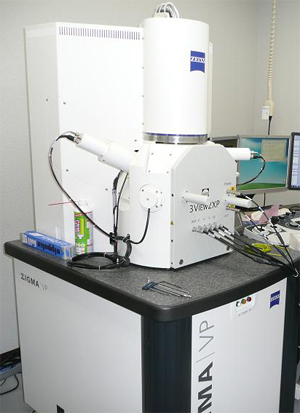
Serial block-face scanning electron microscope(SBF-SEM) is an advanced 3-D imaging equippment. Two different types of SBF-SEM are available; high-resolution type and wide-area type. A resin-embedded biological specimen is trimed by a diamond knife attached inside the chamber, and the block-face images are acquired by scanning electron microscope(SEM) continuously. 3-D structure of the specimen is finally rebuilt from the serial block-face images. 3-D structures of large biological specimens like a brain tissue can be visualized by at dosens of nanometer resolution.
Mutiphoton excitation microscopy
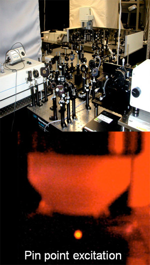
Multi-photon excitation is a method to visualize living tissue by exciting the fluorescence molecules with the tightly focused near-infrared femtosecond pulse laser. Since the longer wavelength is used for multi-photon excitaton, it has a superior deeper tissue penetration and reduced phototoxicity than single-photon excitation. Current projects are the imaging of neurons, glial cells in deep tissues such as mouse brain. Our 2-photon microscope has top level specification for deep tissue imaging. As new project, we recently started to image protein-protein interaction and the activation of signaling molecules by using a 2-photon fluorescence imaging microscope.
A comprehensive behavioral test battery
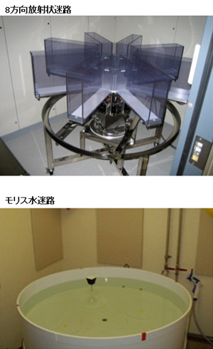
In our laboratory, we can conduct various kinds of behavioral tests for genetically engineered mice, including wire hang, grip strength, light/dark transition, open field, elevated plus maze, hot plate, social interaction, rotarod, prepulse inhibition/startle response, Porsolt forced swim, gait analysis, eight-arm radial maze, T maze, Morris water maze, Barnes maze, object recognition test, cued and contextual fear conditioning, passive avoidance, tail suspension, and 24 hour home cage monitoring.
The primary goal of our research group is to reveal functional significances of genes and their involvement in neuropsychiatric disorders by conducting a comprehensive behavioral test battery on genetically engineered mice.
Analytical equipment for in vivo neuronal, metabolic and physiological parameters in mice and rats
We analyze the following physiological parameters in mice and rats:
1) Single unit record ingfrom motor related brain regions in awake state ,
2) Neurotransmitter release in local brain regions in freemoving animals,
3) Regional neural activity detected as intrinsic signals with taking the advantage of light fluorescent dynamics of flavin or hemoglobin,
4) Energy intake and expenditure in free-moving animals,
5)Body temperature, heart rate and blood pressure in free-moving animals.
