Mechanisms underlying information processing and activity-dependent functional developments in neocortex
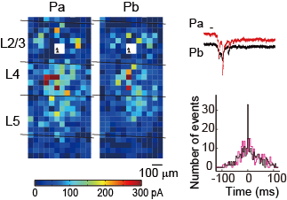
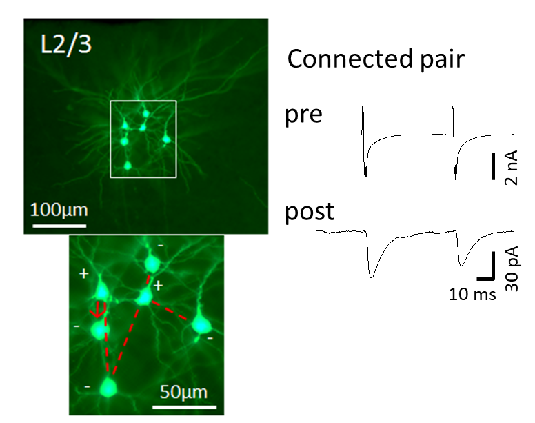
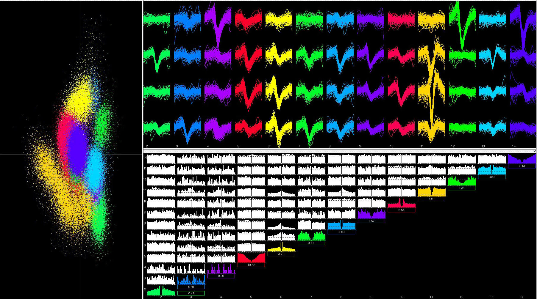
Cortical functions are established and/or modified depending on the postnatal experience and learning. The maturation and improvement of cortical functions may be based on the synaptic plasticity and refinements of neural circuits in the neocortex. To elucidate the mechanisms underlying information processing in sensory cortex and the experience-dependent regulation of that processing, we are studying the relationship between visual functions and the signaling properties of neural circuits using rat and mouse visual cortex. We are also examining the development of neural circuits and functions based on neural activity or synaptic target recognition using specific molecules. To this end, we are analyzing the visual responses of cortical neurons using multi-channel electrodes or calcium imaging with 2-photon microscopy, neural circuit properties with a combination of laser scanning photostimulation and whole-cell recording methods in slice preparations, and neural connections morphologically using modern virus tracers. The following is a list of our main projects currently ongoing.
1. The mechanisms that establish fine-scale networks in visual cortex and the role of these networks in visual information processing
2. Cell-lineage dependent establishment of neural connections and visual responsiveness
3. Synaptic plasticity and visual response plasticity in animals at different developmental stages and in animals subjected to the manipulation of visual inputs during postnatal development
4. Morphological analysis of neural circuits using trans-synaptic virus tracers
5. Neural activities in visual and motor cortex underlying visual cue-triggered behavioral tasks