Revealing the link between eye movements and brain activity
— a comparison of smooth and saccadic visual tracking
2025.10.29
Research
Summary
As the saying goes, "The eyes are the windows to the soul." Our eyes reveal what we feel and help us see the world. We use two eye movements: smooth pursuit, to follow moving objects, and saccades, to quickly shift our gaze. Using functional MRI and the Human Connectome Project’s advanced preprocessing/analysis pipelines, we compared the brain networks supporting these movements. This study, published in Cerebral Cortex, identified both shared and distinct brain activity patterns.Back ground
Smooth pursuit and saccadic eye movements are both essential for vision, yet they serve distinct functions and are controlled by different neural systems. For example, while we can voluntarily make saccades—even with our eyes closed—it is nearly impossible to perform smooth pursuit without visual input. Patients with schizophrenia often show deficits in smooth pursuit, suggesting that these two types of eye movements rely on partly distinct neural networks. However, few studies have directly compared them under equivalent experimental conditions.
Results
Using functional MRI, we examined whole-brain activity during both types of eye movements, carefully matching all parameters except for movement type. By applying the advanced preprocessing and analysis pipelines developed by the Human Connectome Project, we achieved highly precise mapping of brain activity.
Both movements, compared with fixation, activated a broad, overlapping network including early and higher visual areas in the occipital lobe, regions along the intraparietal sulcus in the parietal lobe, several oculomotor areas in the frontal lobe, the cerebellum, and subcortical regions. Interestingly, both also showed similar deactivation patterns in certain brain regions during eye movements. At the same time, distinct patterns emerged: smooth pursuit elicited stronger activity in visual and parietal regions, the posterior cingulate sulcus, and the superior parietal lobule, whereas saccades more strongly engaged frontal oculomotor areas and parietal regions along the intraparietal sulcus.
These findings provide new insight into the shared and distinct neural architectures supporting human eye movements. They may also enhance our understanding of psychiatric and neurological disorders such as schizophrenia and help identify early diagnostic markers. Beyond medicine, individual differences in pursuit ability could also relate to ball-sport performance, suggesting future applications in sports science. Our eye movements, through which we perceive the outside world, may serve as a unique window into the workings of the human mind and brain.
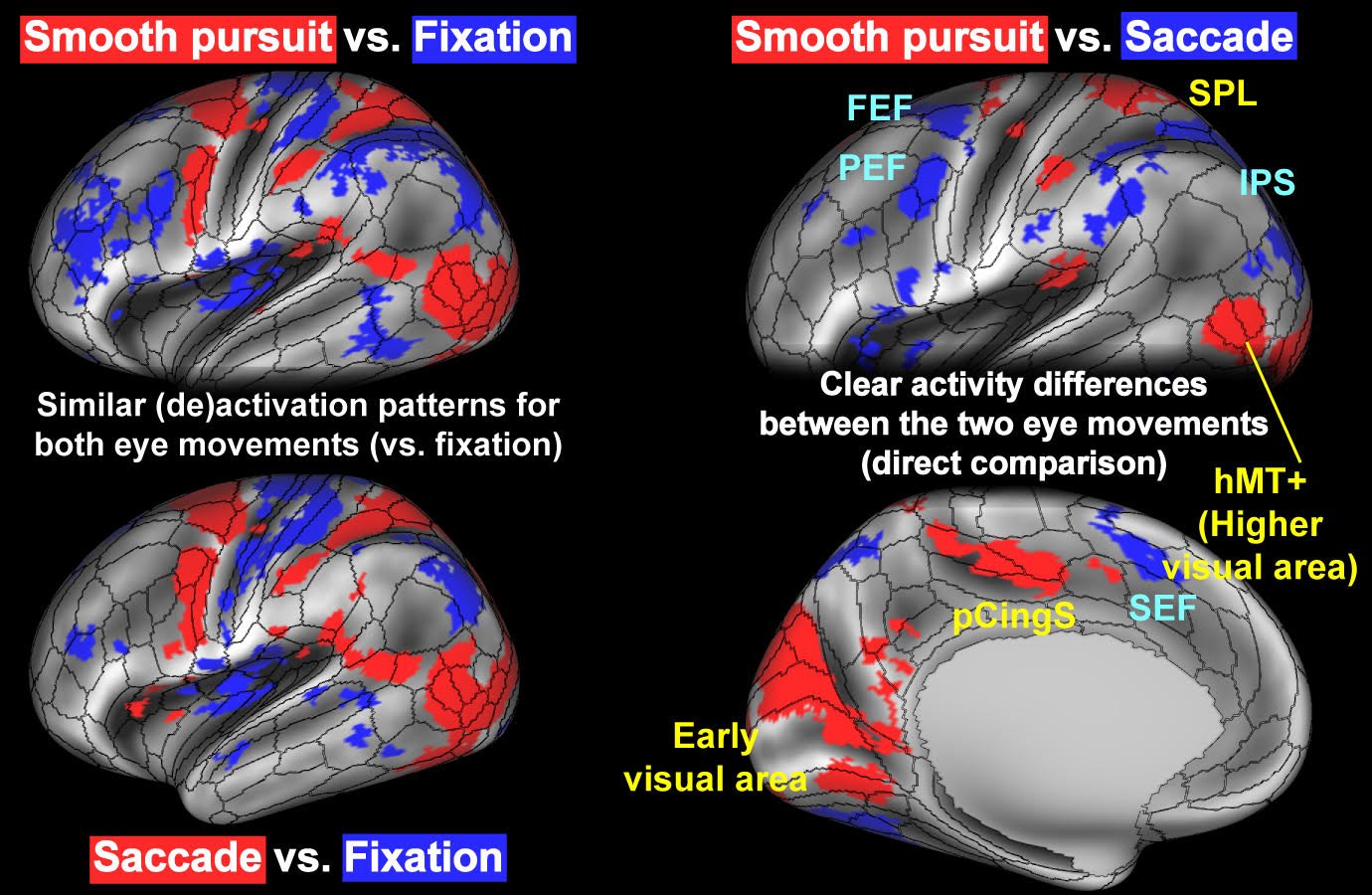 When compared with fixation, both smooth pursuit (upper left) and saccadic (lower left) eye movements showed similar patterns of brain activation (red) and deactivation (blue). In contrast, a direct comparison between the two (right panels) revealed distinct regions more strongly associated with smooth pursuit (red) and with saccades (blue). Abbreviations: FEF, frontal eye field; hMT+, human middle temporal complex; IPS, intraparietal sulcus; PEF, premotor eye field; pCingS, posterior cingulate sulcus; SEF, supplementary eye field; SPL, superior parietal lobule.
Research member and collaborative researcher
Tetsuya Yamamoto (Section of Brain Function Information, National Institute for Physiology; Core for Spin Life Sciences, Okazaki Collaborative Platform, National Institutes of Natural Sciences)
Kenichiro Miura (Section of Brain Function Information, National Institute for Physiology; Department of Pathology of Mental Diseases, National Institute of Mental Health, National Center of Neurology and Psychiatry; Institute for the Advanced Study of Human Biology, Kyoto University)
Keiji Matsuda (Human Informatics and Interaction Research Institute, National Institute of Advanced Industrial Science and Technology)
Junya Matsumoto (Department of Pathology of Mental Diseases, National Institute of Mental Health, National Center of Neurology and Psychiatry)
Ryota Hashimoto (Department of Pathology of Mental Diseases, National Institute of Mental Health, National Center of Neurology and Psychiatry)
Seiji Ono (Institute of Health and Sport Sciences, University of Tsukuba)
Norihiro Sadato (Section of Brain Function Information, National Institute for Physiology; Core for Spin Life Sciences, Okazaki Collaborative Platform, National Institutes of Natural Sciences; Research Organization of Science and Technology, Ritsumeikan University)
Masaki Fukunaga (Section of Brain Function Information, National Institute for Physiology; Core for Spin Life Sciences, Okazaki Collaborative Platform, National Institutes of Natural Sciences; Physiological Science Program, Graduate Institute for Advanced Studies, SOKENDAI)
Funding
This study was supported by JSPS KAKENHI Grant Number 20K06920 (K.M.), 22K12809 (T.Y.), 23K24749 (S.O.), 23K25200 (M.F.), and 24H00622(N.S.), Brain/MINDS 2.0 from AMED JP23wm0625001 (M.F.), and MEXT/CURE JPMXP1323015488 (Spin-L program No, spin24XN038) (K.M., S.O.).Journal article
Title: Brain-wide activation and deactivation maps during smooth and saccadic tracking in humansAuthors: Tetsuya Yamamoto, Kenichiro Miura, Keiji Matsuda, Ryota Hashimoto, Seiji Ono, Norihiro Sadato, Masaki Fukunaga
Journal: Cerebral Cortex
Issue: 35(8)
Date: 2025/9/10
URL (abstract): https://academic.oup.com/cercor/article/35/8/bhaf242/8250586
DOI: 10.1093/cercor/bhaf242