How does vision suppress the auditory cortex?
Ultra-High-Field fMRI Provides Detailed Mapping of Interhemispheric Mechanisms in Cross-Modal Sensory Suppression
2026.03.24
Research
Summary
When we experience the world, our senses rarely work in isolation. Visual, auditory, and tactile input constantly interacts in the brain. While previous functional MRI studies have shown that activity in one sensory system can suppress another, the specific mechanism underlying this phenomenon remains under investigation. Researchers at the National Institute for Physiological Sciences used ultra-high-field (7-Tesla) functional magnetic resonance imaging (fMRI) to further characterize and validate how the human brain coordinates sensory inputs from multiple modalities.Results
The team utilized the brain’s contralateral processing, in which visual information from one field is processed by the opposite hemisphere. By presenting visual stimuli to only the left or right side of the screen, the researchers could isolate the visual cortex activity to one hemisphere. This allowed them to test whether the resulting suppression in the auditory cortex was a local effect or a broader network response.
“Our goal was to understand where cross-modal suppression happens and what kind of neural mechanisms might underlie it,” says lead author Toshikazu Miyata. “By using lateralized visual stimuli, we could directly test whether these effects depend on the activity of the visual cortex in the same hemisphere.”
The researchers found a clear result regarding the activity within the primary auditory cortex. When participants viewed visual stimuli, activity in the auditory cortex decreased in both hemispheres, regardless of which visual field was stimulated.
“This bilateral pattern, confirmed through individual-level mapping, points to robust interhemispheric suppression mechanisms,” explains senior author Hiromasa Takemura. “It suggests that visual input reduces auditory cortex activity through broad coordination rather than local interactions.”
In contrast, no such cross-modal suppression was observed in the sensory thalamus, a key early processing hub for visual and auditory information. This indicates that suppressions are more likely to emerge at the level of the cerebral cortex rather than at earlier subcortical stages.
Together, the findings refine our understanding of how the human brain coordinates multiple senses. Rather than being a passive consequence of blood flow changes, cross-modal suppression in the auditory cortex appears to reflect active neural regulation across hemispheres.
“Our findings reinforce and refine our understanding of sensory interaction,” says Miyata. “By confirming the cortical nature and bilateral spread of this response using high-precision mapping, we provide a more robust foundation for understanding how the healthy adult brain maintains sensory balance.”
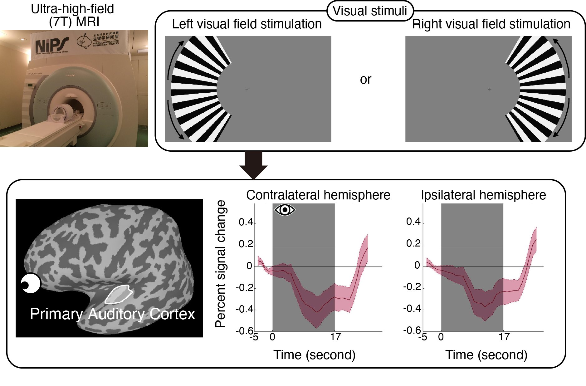
Figure. Upper left: Ultra-high field (7 Tesla) MRI scanner at the National Institute for Physiological Sciences. Upper right: Visual stimuli used in the fMRI experiment. Moving gratings were presented to either the left or right visual field. Lower left: Location of the primary auditory cortex in the human brain. Lower right: Brain activity in the contralateral and ipsilateral primary auditory cortex during visual stimulation. The horizontal axis represents time, and the vertical axis represents the percent signal change in brain activity. The onset of visual stimulation is at 0 seconds, with the gray shaded area indicating the stimulus duration. The red line represents the mean across all participants, and the magenta shaded area represents ±1 standard error (SE) from the mean. Comparable levels of brain activity suppression were observed in both hemispheres.
Research member and collaborative researcher
Toshikazu Miyata (National Institute for Physiological Sciences; Department of Quantitative and Imaging Biology, Headquarters for Co-Creation Strategy, National Institutes of Natural Sciences; Princeton University)
Masaki Fukunaga (National Institute for Physiological Sciences; SOKENDAI; Core for Spin Life Sciences, Okazaki Collaborative Platform)
Junxiang Luo (National Institute for Physiological Sciences; SOKENDAI; Core for Spin Life Sciences, Okazaki Collaborative Platform)
Isao Yokoi (National Institute for Physiological Sciences)
Tetsuya Yamamoto (National Institute for Physiological Sciences; SOKENDAI; Core for Spin Life Sciences, Okazaki Collaborative Platform)
Ayumi Yoshioka (National Institute for Physiological Sciences; Ritsumeikan University)
Jiajia Yang (Okayama University)
Tomoyo Morita (National Institute of Information and Communications Technology; Osaka University)
Hiromasa Takemura (National Institute for Physiological Sciences; Department of Quantitative and Imaging Biology, Headquarters for Co-Creation Strategy, National Institutes of Natural Sciences; SOKENDAI; Core for Spin Life Sciences, Okazaki Collaborative Platform)
Funding
The study was supported by the Grants-in-Aid for Japan Society for the Promotion of Science (KAKENHI), the Joint Research Program of the National Institute for Physiological Sciences, and the MEXT Promotion of Development of a Joint Usage/Research System Project: Coalition of Universities of Research Excellence Program (CURE).Journal article
Title: Characteristics of cross-modal negative BOLD responses in the human sensory subcortex and cortex
Authors: Toshikazu Miyata, Masaki Fukunaga, Junxiang Luo, Isao Yokoi, Tetsuya Yamamoto, Ayumi Yoshioka, Jiajia Yang, Tomoyo Morita, Hiromasa Takemura
Journal: Journal of Neurophysiology, Volume 135, Issue 4, Page 747–759
Date: 2026/02/05
URL (abstract): https://journals.physiology.org/doi/full/10.1152/jn.00396.2025