



Brain reorganization after spinal cord injury revealed by stimulation of the non-paralyzed hand and its relationship to motor recovery
2026.06.05
Research
Summary
Our study has uncovered a key feature of sensorimotor system reorganization during recovery from spinal cord injury. By combining longitudinal functional MRI to track whole-brain activity and connectivity with behavioral assessments of grasping movements in monkeys with unilateral spinal cord injury, we found that changes in brain responsiveness to non-paralyzed hand stimulation may reflect sensorimotor system reorganization during recovery. These findings were published in the international journal Scientific Reports.
Results
The brain processes sensory inputs from the hands. However, when one side of the spinal cord is damaged, it remains unclear how this information processing changes and whether this change contributes to recovery of hand movement.
To investigate this, we conducted intensive longitudinal functional MRI, combined with behavioral assessments of grasping movements, in five monkeys with unilateral spinal cord injury.
In the four monkeys that showed good recovery, responses in bilateral sensorimotor hand areas to stimulation of the severely paralyzed hand changed little. In contrast, responses to stimulation of the non-paralyzed hand increased 2–5 weeks after injury and then gradually declined over time. These changes were associated with the reorganization of motor networks across the brain, including the premotor and supplementary motor areas. In addition, the sensorimotor cortex ipsilateral to the injury exhibited stronger responses that correlated inversely with grasping performance. This indicates the relationship between stronger response and poorer grasping performance. In fact, the monkey with poor recovery showed widespread and persistent responses correlated inversely with grasping performance.
Together, these findings suggest that early increases in brain responsiveness reflect a reduction of inhibitory control (disinhibition), which may facilitate large-scale reorganization of motor networks. The subsequent reduction of this disinhibition leads to long-term recovery after spinal cord injury in primates.
By examining brain responses to stimulation of the non-paralyzed hand, we identified patterns of sensorimotor reorganization during recovery of hand movement after spinal cord injury. This study differs from previous work centered primarily on the paralyzed hand, by revealing mechanisms of brain adaptation after spinal cord injury from a new perspective based on responses to stimulation of the non-paralyzed hand. These findings provide new insight into patterns of brain reorganization after partial spinal cord injury and can contribute to the development of more effective rehabilitation strategies in the future.
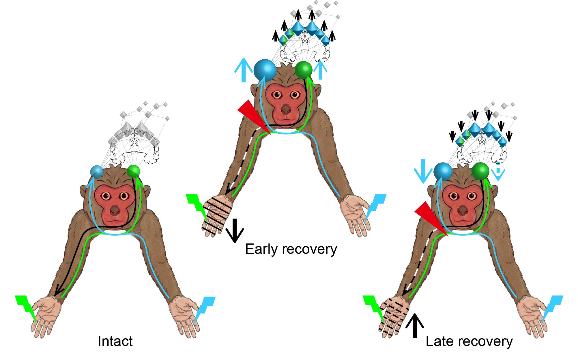
These figures show brain responses to tactile stimulation of the hand and associated motor networks before injury (intact), and during early and late recovery periods after injury. In the intact state, the sensorimotor hand areas (light blue and green spheres) receive sensory inputs from, and send motor outputs (black arrow shown for one hemisphere) to, the opposite hand. In the early recovery period after unilateral spinal cord injury (red triangle), the hand ipsilateral to the injury was paralyzed, indicated by oblique lines on the hand. Bilateral sensorimotor hand areas exhibited increased responses to stimulation of the non-paralyzed hand, indicated by blue upward arrows beside spheres. These responses were associated with increased involvement of motor networks across the brain, including the premotor (small bicones) and supplementary motor areas (large bicones). Paired associations are shown in light blue or green. In addition, stronger responses in the sensorimotor region were associated with poorer grasping performance, indicated by a black downward arrow beside the hand. In the late recovery period, these increased responses declined as recovery progressed, in association with changes in motor network involvement, as shown by the upward and downward arrows.
Collaborative Researcher
Takamichi Tohyama (Section of Brain Function Information, National Institute for Physiological Sciences; Department of Rehabilitation Medicine, School of Medicine, Fujita Health University)
Reona Yamaguchi (Institute for the Advanced Study of Human Biology [WPI-ASHBi], Kyoto University; Department of Neuroscience, Graduate School of Medicine, Kyoto University)
Naokazu Goda (Section of Brain Function Information, National Institute for Physiological Sciences; Core for Spin Life Sciences, Okazaki Collaborative Platform, National Institutes of Natural Sciences; Physiological Science Program, Graduate Institute for Advanced Studies, SOKENDAI)
Tetsuya Yamamoto (Section of Brain Function Information, National Institute for Physiological Sciences; Core for Spin Life Sciences, Okazaki Collaborative Platform, National Institutes of Natural Sciences; Research Organization of Science and Technology, Ritsumeikan University)
Norihiro Sadato (Core for Spin Life Sciences, Okazaki Collaborative Platform, National Institutes of Natural Sciences; Research Organization of Science and Technology, Ritsumeikan University; Center for Research Collaboration, National Institute for Physiological Sciences)
Tadashi Isa (Institute for the Advanced Study of Human Biology [WPI-ASHBi], Kyoto University; Department of Neuroscience, Graduate School of Medicine, Kyoto University; Core for Spin Life Sciences, Okazaki Collaborative Platform, National Institutes of Natural Sciences; Section of Advanced Project Promotion, National Institute for Physiological Sciences)
Masaki Fukunaga (Section of Brain Function Information, National Institute for Physiological Sciences; Core for Spin Life Sciences, Okazaki Collaborative Platform, National Institutes of Natural Sciences; Physiological Science Program, Graduate Institute for Advanced Studies, SOKENDAI)
Reona Yamaguchi (Institute for the Advanced Study of Human Biology [WPI-ASHBi], Kyoto University; Department of Neuroscience, Graduate School of Medicine, Kyoto University)
Naokazu Goda (Section of Brain Function Information, National Institute for Physiological Sciences; Core for Spin Life Sciences, Okazaki Collaborative Platform, National Institutes of Natural Sciences; Physiological Science Program, Graduate Institute for Advanced Studies, SOKENDAI)
Tetsuya Yamamoto (Section of Brain Function Information, National Institute for Physiological Sciences; Core for Spin Life Sciences, Okazaki Collaborative Platform, National Institutes of Natural Sciences; Research Organization of Science and Technology, Ritsumeikan University)
Norihiro Sadato (Core for Spin Life Sciences, Okazaki Collaborative Platform, National Institutes of Natural Sciences; Research Organization of Science and Technology, Ritsumeikan University; Center for Research Collaboration, National Institute for Physiological Sciences)
Tadashi Isa (Institute for the Advanced Study of Human Biology [WPI-ASHBi], Kyoto University; Department of Neuroscience, Graduate School of Medicine, Kyoto University; Core for Spin Life Sciences, Okazaki Collaborative Platform, National Institutes of Natural Sciences; Section of Advanced Project Promotion, National Institute for Physiological Sciences)
Masaki Fukunaga (Section of Brain Function Information, National Institute for Physiological Sciences; Core for Spin Life Sciences, Okazaki Collaborative Platform, National Institutes of Natural Sciences; Physiological Science Program, Graduate Institute for Advanced Studies, SOKENDAI)
Funding
This study was supported by the Strategic Research Program for Brain Sciences from AMED (JP17dm0107118), Brain/MINDS 2.0 from AMED (JP24wm0625001) and KAKENHI (JP19K22985, JP23K25200) of Japan to M. F., Brain/MINDS-beyond from AMED (JP18dm0307005) to N. S. and T. I., and KAKENHI of Japan (JP22K15624) to T. T., and KAKENHI of Japan (JP22H04992) to T. I.
Journal article
Title: Enhanced sensorimotor cortex responsiveness to nonplegic hand stimulation and motor network assembly during recovery after spinal cord injury in primates
Authors: Takamichi Tohyama, Reona Yamaguchi, Naokazu Goda, Tetsuya Yamamoto, Norihiro Sadato, Tadashi Isa and Masaki Fukunaga
Journal: Scientific Reports
Date: 27 May 2026
URL (abstract): https://www.nature.com/articles/s41598-026-54528-7