生理学実験技術データベース
Experimental Techniques for Physiological Sciences
G3-19
Last update: 2010-03-30
MRI撮影用頭部固定具
Head fixture for MRI photography
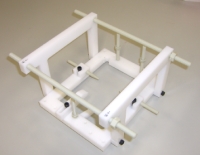
| Summary | In the highly advanced brain function study that I used a monkey for, it is very important that I know shape and the state of the brain before an operation. I confirm yes or no of the damage of the brain beforehand, and the reason is because it locates the electrode to install. I get the form image of the monkey brain with an MRI device, but the head has to fix it to know the coordinate position of the brain definitely not to move. I produced the device which could fix the head relatively easily this time. |
|---|---|
| 用途 | MRI装置でサルの頭部撮影時に使用する。 |
| 特徴 |
バードケージコイル、サーフェースコイルにも対応できる。 通常固定時に水平を取るため水準器を用いるが、この固定具で水平になるため水準器の必要が無い。 外耳孔の固定具全体が容易に着脱可能で、イヤーバーもねじ式で、従来より微調節がし易い構造となっている。 生理研に導入されているシーメンス社製3TMRI装置用 |
|
構成 使用方法 |
製作した固定具の構成は、中心となるフレーム部、外耳孔、眼窩下縁、上顎部の固定具、MRI装置のベットに設置するためのウイングや脚、サーフェースコイル取付け具からなっている。 頭部固定時にはフレーム部に脚を付けて安定に作業を行える。撮影に際しては、サーフェイスコイル使用時は、ウイングを取付けて、MRI装置ベットに乗せるような構造にし、バードケージコイル使用時は、脚を上向き方向に取付け、バードケージコイルの枠かた吊り下げる構造になっている。 |
| 詳細資料 | 外観、使用状況(バードケージ、サーフェース) |
| その他 | MRI撮影データ(所外非公開) |