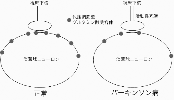
パーキンソン病の際、視床下核から淡蒼球へのグルタミン酸作動性投射の活動性が亢進する事が、症状の発現に重要な役割を果たすと考えられている。本研究においては、霊長類を用いてパーキンソン病の際の代謝調節型グルタミン酸受容体(mGluRs)の活性について調べた。MPTPを投与して作成したパーキンソン病モデル動物においては、mGluRsのうちmGluR1αの発現のみが淡蒼球外節・内節で減少していた。さらに、この機能を調べるために、mGluR1の作動薬を正常な霊長類の淡蒼球ニューロンに作用させたところ興奮性作用を示し、逆に拮抗薬を投与すると抑制されることがわかった。さらに、パーキンソン病モデル動物においては、このような薬物の作用が減弱していることが示された。以上の結果は、パーキンソン病では淡蒼球における代謝調節型グルタミン酸受容体の発現が減少し、これは視床下核-淡蒼球投射の過剰興奮に対する代償作用を担っている可能性を示唆する(図参照)。
Kaneda K, Tachibana Y, Imanishi M, Kita H, Shigemoto R, NambuA & Takada M (2005) Downregulation of metabotropic glutamate receptor 1a in globus pallidus and substantia nigra of parkinsonian monkeys. Eur J Neurosci. Dec;22(12):3241-54.

Enhanced glutamatergic neurotransmission via the subthalamopallidal or subthalamonigral projection seems crucial for developing parkinsonian motor signs. In the present study, the possible changes in metabotropic glutamate receptors (mGluRs) expression were examined in the basal ganglia of a primate model for Parkinson's disease. When the patterns of immunohistochemical localization of mGluRs in monkeys administered systemically with 1-methyl-4-phenyl-1,2,3,6-tetrahydropyridine (MPTP) were analyzed in comparison with normal controls, we found that expression of mGluR1, but not of other subtypes, was significantly reduced in the internal and external segments of the globus pallidus and the substantia nigra pars reticulata. To elucidate the functional role of mGluR1 in the control of pallidal neuron activity, extracellular unit recordings combined with intrapallidal microinjections of mGluR1-related agents were then performed in normal and parkinsonian monkeys. In normal awake conditions, the spontaneous firing rates of neurons in the pallidal complex were increased by DHPG, a selective agonist of group I mGluRs, whereas they were decreased by AIDA, a selective antagonist of group I mGluRs or LY367385, a selective antagonist of mGluR1. These electrophysiological data strongly indicate that the excitatory mechanism of pallidal neurons by glutamate is mediated at least partly through mGluR1. The effects of the mGluR1-related agents on neuronal firing in the internal pallidal segment became rather obscure after MPTP treatment. Our results suggest that the specific downregulation of pallidal and nigral mGluR1in the parkinsonian state may exert a compensatory action to reverse the overactivity of the subthalamic nucleus-derived glutamatergic input that is generated in the disease.