[A-1]
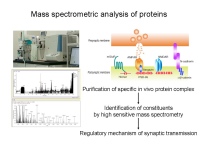
Mass spectrometric analysis of proteins
(Div of Membrane Physiology: Fukata Lab)
Determining the native protein complex is essential for understanding the physiological and pathological mechanisms of living organisms. We have recently established relatively simple purification method, which allows high recovery and specific isolation of the protein complex in vivo. Combined with mass spectrometry, we have identified various in vivo protein complexes (or protein networks). In this course, we will perform a series of experiments for the identification of in vivo protein complex.
[A-2] in situ hybridization
(Div of Neurobiology and Bioinformatics: Ikenaka Lab)
Histochemical analysis of mRNA localization is a powerful tool to study the
function of a gene. Especially in the brain it is essential to use in situ hybridization technique to identify the cell type expressing the gene because cell types present in the brain is so complex. We will analyze astrocyte specific and oligodendrocyte specific gene expression by the in situ hybridization.
[A-3]
Quantitative electron microscopic analysis of synapses
(Div of Cerebral Circuitry: Kawaguchi Lab)
Experience of electron microscopy is required.
Quantitative analysis of synaptic structures and distributions is importantfor neural circuit understanding. One morphological tool for this purpose is three dimensional reconstruction of synapses and surround structures from the serial ultrathin sections. In this course, we introduce this technique using the prevailing free software "Reconstruct" and show the way to obtain quantitative parameters (the synapse density, and surface/cross sectional area of dendrites/somata) from the 3D image.
crick to enlarge
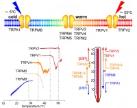
[A-4] Biosensor molecules for pain and temperature
(Div of Cell Signalling: Tominaga Lab)
There are nine thermosensitive TRP channels expressed in sensory neurons and other cells some of which are known to be also sensitive to nociceptive stimuli. Simultaneous recordings of the temperature-change-evoked current responses (in either whole-cell recordings or single-channel recordings from the excised membrane patches) and bath temperature would tell us that the thermosensitive TRP channels have distinct temperature threshold for their activation. An arrhenius plot provides much more price temperature threshold.
crick to enlarge
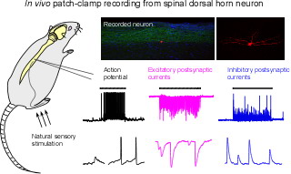
[A-5] Patch clamp recording in vivo
(Div of Neural Signaling: Imoto/Furue Lab)
Patch-clamp recording technique has been mainly applied to in vitro preparations such as culture cells, dissociated cells and brain slices, contributing greatly to understanding ionic mechanisms of channels/receptors, and synaptic transmission in the neuronal circuits. To further elucidate the physiological significance of the neuronal and synaptic activities observed in such in vitro preparations, we have recently developed an in vivo patch-clamp recording technique from spinal dorsal horn neurons. In this course, we will show in vivo patch-clamp recordings from spinal superficial dorsal horn neurons, their synaptic responses to natural cutaneous noxious stimulation and the antinociceptive action of anesthetic drugs.
crick to enlarge
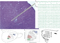
[A-6] Multichannel recording in anesthetized animals
(Div of Behavioral Development: Isa Lab)
Multichannel recording provides by far richer information than recording wiith a single channel electrode for understanding the information processing in the brain.
In this course, we wil try multichannel recordings of single units, field potential together with recording of electrocorticogram(ECoG) in the barrel cortex of anesthetized rats.
Trainees will learn how to conduct appropriate electrophysiological experiments in anesthetized animals and how to analyze the data.
[A-7] Electroencephalography (EEG) and magnetic electroencephalography
(Div of Sensori-Motor Integration: Kakigi Lab)
We investigate human brain functions non-invasively mainly using magnetoencephalography (MEG) and electroencephalography (EEG). The following investigations are in progress at present.
Sensory system: By recording brain responses to visual, auditory, somatosensory or pain stimuli, the organization of sensory processing in the human brain is being investigated.
Even-related brain responses: Using various psychophysical tasks or paradigms, we are investigating cognitive processing of the brain (higher brain functions).
Application of brain research to education and society: Recently we focused on the development of brain function in infants and children. EEG and near-infrared spectroscopy (NIRS) are useful in this study, since these methods can be applied to infants and children who can not hold their heads still for a long time.
Group B 2nd week
[B-1] Phase-contrast cryo-electron microscopy (Div of Nano-Structure Physiology: Nagayama Lab)
This course is aimed at introducing beginner to intermediate level electron microscopists to the methods and techniques of cryo-electron microscopy of biological specimens. In addition the students will have the chance to learn first-hand the novel methods of Zernike phase contrast and its application to electron cryo-tomography of ice-embedded biological specimens. The Zernike phase contrast technique for electron microscopy was firstly developed and applied in our laboratory and is gradually gaining popularity worldwide.
The course will focus mostly on the experimental aspects of phase contrast cryo-electron microscopy with a short introduction to the theoretical basis. The participants will first learn how to prepare cryo-specimens of proteins and other small biological objects by cryo-plunging. Then they will be introduced to the low dose operation, phase plate manipulation and tomography data collection by a 200 kV cryo-electron microscope. Finally, they will use the collected data to generate a 3D tomogram of the investigated specimen by tomography reconstruction software.
[B-2] SDS Freeze-fracture replica labeling electron microscopy
(Div of Cerebral Structure: Shigemoto Lab)
Experience of electron microscopy is required.
In this Laboratory experience, we will show a series of operations for preparation of SDS-FRL materials and observe replicas from the hippocampus and cerebellum labeled for AMPA-type and NMDA-type glutamate receptors. You can learn advantages and disadvantages of this technique and how to interpret the results on replica morphology and molecular localization in the brain.
crick to enlarge
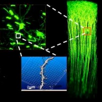
[B-3] Multiphoton confocal microscopy
(Div of Homeostatic Development: Nabekura Lab)
Application of non-linear 2 photon laser scanning microscopy (TPLM) to living animals enables us to obtain 4D imaging of various fine structures with submicron resolution in deep tissues, such as brain. Using this advanced imaging method, we have studied the remodeling of neuronal circuits in development and repair, the interaction between glia and neurons, the migration of GABAergic neurons and the dynamics of cortical neuronal activities in various pathological conditions.
In this course, we would like to show:
how to make a cranial window for imaging of mouse cortex
the application of TPLM to visualize the pyramidal neurons, their dendrites and spines expressing GFP in living mice.
crick to enlarge
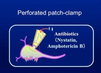
[B-4] Perforated patch recordings
(Div of Homeositatic Development: Nabekura Lab)
The advent of the patch-clamp technique has revolutionized to the study of membrane physiology. However, the whole-cell recording suffers from drawbacks related to the exposure of intracellular space to artificial solutions in the recording pipette. The dialysis of the cell interior with pipette solution washes out cytoplasmic biochemicals which is required for channel activities and second messenger-mediated responses. Perforated patch recording was devised to gain electrical access to the cell interior without diffusion of cellular constituents into patch pipette. This technique used the antibiotics, such as nystatin and gramicidin, to form small pores in the membrane under the patch pipette. These pores allow only monovalent ions to pass but prevent the movement of larger molecules. Thus, one has electrical access to the entire cell with minimal dialysis of the cytoplasmic substances.
crick to enlarge
[B-5] Laser scanning photostimulation of brain slices
(Div of Developmental Neurophysiology: Yoshimura Lab)
We have been investigating functional neocortical circuits using laser scanning photostimulation, which allows us to rapidly map synaptic connections in acute brain slices. In this Laboratory experience, we will show a series of operations to map the location of neurons presynaptic to whole-cell recorded neurons by focal activation of the former neurons using uncaged glutamate photostimulation in rat visual cortical slices.
[B-6] Neural recordings in behaving animals
(Div of System Neurophysiology: Nambu Lab)
Recording neuronal activity from behaving animals is a powerful technique to understand how neuronal circuitry works in the living organisms. This technique has been developed originally using primates, but is also applicable to rodents, especially transgenic animals. Using this technique, we can avoid effects of anesthetics and use the same animals repeatedly. In addition, we can utilize electrical stimulation and local drug injections. In this course, basic techniques, such as animal operation, making metal electrodes and recording neuronal activity from awake animals, will be trained for rookies that just started or will soon start these experiments.
crick to enlarge
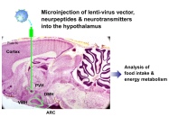
[B-7] Analysis of feeding behavior and peripheral mechanism
(Div of Endocrinology and Metabolism: Minokoshi Lab)
Within the central nervous system, the hypothalamus is a critical center that regulates the homeostatic activities by integrating autonomic nervous system, endocrine system and immune function. In this school, we will show how to study the mechanism of food intake and metabolic regulation in the hypothalamus. Especially, you can learn the microinjection method of neuropeptides into the brain in mice and analysis of food intake and water drinking behavior.
[B-8] Magnetic resonance imaging of brain function
(Div of Cerebral Integration: Sadato Lab)
The goal of Division of Cerebral Integration is to understand the physiology of human voluntary movement and other mental processing including language using noninvasive functional neuroimaging technique, mainly fMRI. In particular, understanding of the mechanisms of plastic change in the human brain accompanied by learning, sensory deafferentation, and development is the research target. Recently, we have been focusing on the development of social cognition. Multimodality approach including EEG, MEG, and NIR is considered when
appropriate.
Explanation of the Figure: Brain areas commonly activated by social and monetary rewards. Why are we nice to others? One answer provided by social psychologists is because it pays off. A social psychological theory stated that we do something nice to others for a good reputation or social approval just like we work for salary. Although this theory assumed that social
reward of a good reputation has the same reward value as money, it was unknown whether it recruits the same reward circuitry as money in human brain. In this study, we found neural evidence that perceiving one's good reputation formed by others activated the striatum, the brain's reward system, in a similar manner to monetary reward. Considering a pivotal role played by a good reputation in social interactions, this study provides an important first step toward neural explanation for our everyday social behaviors.
Copyright (C) 2009 National Institute for Physiological Sciences, All right reserved.