Cryo-EM structure of a Marseilleviridae virus particle reveals a large internal microassembly
Giant virus is a virus that is larger than mycoplasma (0.2 nm *1), which is the smallest organism. Since the giant virus shows many structural characteristics closer to cellular organisms, it is drawing attention as being present at the boundary between virus and cellular organism.
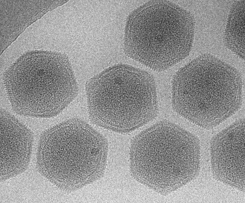
In this study, our collaborative research group revealed the detailed structure of Melbourne virus (MelV), one of the newly discovered giant viruses, using cryo-electron microscopy (cryo-EM). MelV is a giant virus found in the freshwater pond in Melbourne and was classified as Marseilleviridae family, a new family of giant viruses. We found that the diameter of the virus is about 250 nm, and the particle is covered with an icosahedral capsid consisted of T = 309. In the capsid, there was a lipid bilayer fixed at the apex of the capsid, and the viral genome was wrapped in it. The most characteristic of MelV was that the virus particle included one high-density granule with a diameter of about 30 nm, which we named LDB. Deduced from its density, LDB was predicted to be a nuclear protein.
From this study, we were able to add one new structural knowledge of the giant virus.
*1 1 nm is equal to 1/1000,000 mm.
Collaborative Researcher
Kenta Okamoto (Uppsala University, Sweden)
Funding
The Swedish Research Council; The Knut and Alice Wallenberg Foundation; The European Research Council; The Rontgen-Angstrom Cluster; The Swedish Foundation for International Cooperation in Research and Higher Education (STINT); The European Regional Development Fund at the European Extreme Light Infrastructure; KAKENHI from the Ministry of Education, Culture, Sports, Science and Technology (MEXT) of Japan; A Grant-in-Aid for Scientific Research on Innovation Area from MEXT of Japan; The Collaborative Study Program of National Institute for Physiological Sciences; The CNRS, Aix-Marseille University and the French National Research Agency