Marseilleviruses newly isolated in Japan reveal the local diversity of the giant virus family
In 2003, the bacteria had been called “Bradford coccus” turned out to be a giant virus, and it surprised people. Since then, viruses, which are larger than the smallest unicellular organisms, have been found around the world, and these have become called "giant viruses". Giant viruses exist by using a single cell organism such as amoeba as host. Several giant viruses have been found in Japan.
In this study, a research team led by Professor Masaharu Takemura in Tokyo University of Science identified the cytopathic effect of amoeba, which indicates the presence of a giant virus, in water samples collected at three watersides in Japan. Genomic analysis of the supernatant predicted the existence of the giant virus, Marseilleviridae. In collaboration study with NIPS, we examined the amoeba cells showing this cytopathic effect with an electron microscope. We were able to identify a large number of icosahedral large viruses with a diameter of about 250 nm inside the cells (Figure). Furthermore, a special organ called "Virion Factory" was formed in the cytoplasm, which produce the viruses. These characteristics confirmed that this is a Marseilleviridae. From the places where the samples were collected, viruses was named "Hokutovirus" "Kashiwazakivirus" "Kyotovirus" of Marseilleviridae, respectively.
So far, it has normally been identified only one giant virus from a single location. However, in this study, the viruses having different types of major capsid protein (MCP) genes were intriguingly identified from the same water samples collected from the three locations. In contrast, viruses having the same MCP gene was found from the different sampling locations.
This result reverses the conventional wisdom and shows for the first time that giant viruses are distributed widely and diversely. In addition, it gives important knowledge in the evolution of the giant viruses. Further research will clarify the relationship between the giant viruses and cellular organisms in nature in the future.
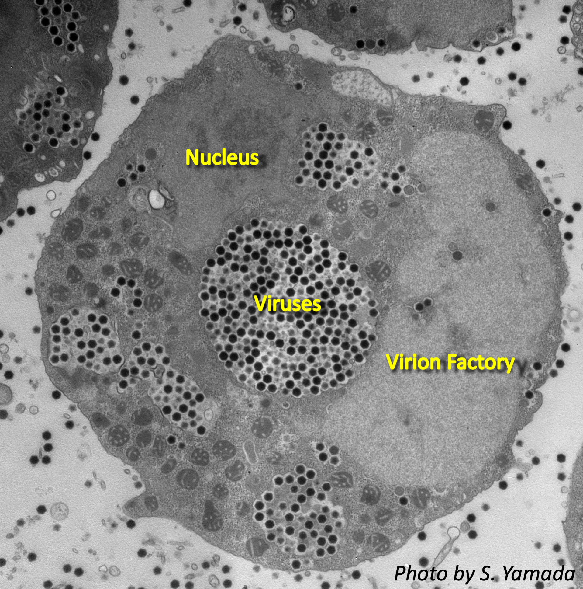
Figure 1 Electron microscope image of amoeba infected with Kashiwazakivirus.
Collaborative Researcher
Masaharu Takemura (Tokyo University of Science)
Kazuyoshi Murata (NIPS)
Funding
KAKENHI (JSPS), NIPS Collaborative Study Program
Release Source
Title: Fifteen Marseilleviruses Newly Isolated From Three Water Samples in Japan Reveal Local Diversity of Marseilleviridae
Authors: Keita Aoki, Reika Hagiwara, Motohiro Akashi, Kenta Sasaki, Kazuyoshi Murata, Hiroyuki Ogata and Masaharu Takemura
Journal: Frontiers in Microbiology
Issue: 10, 1152
Date: 2019, May 24 Published
URL: https://doi.org/10.3389/fmicb.2019.01152
DOI: 10.3390/ijms20092308.