Structural changes in the large capsid during particle formation of the giant virus Medusavirus
2024.11.18
Research
Viruses larger than small bacteria (approximately 200 nm in diameter*1) are generally called giant viruses. Some giant viruses are covered with a capsid (shell) that has a geometric icosahedral (soccer ball) structure regardless of their size, and although there are various theories as to how such giant virus particles are formed, it is currently not well understood. Medusavirus (1), discovered by our collaborative research group and reported in 2019, is one of the giant viruses with a regular icosahedral structure, and interestingly, we found that amoebas infected with this virus simultaneously release a large number of immature particles in addition to mature virus particles (2). In this study, we used a cryo-electron microscopy*2 to examine the structures of mature and immature Medusavirus particles (Figure 1A), and clarified the structural changes of the capsid during the Medusavirus particle formation process (Figures 1B-D).In Medusavirus, a particle consisting of only the capsid is formed first, followed by the formation of a lipid membrane inside that later covers the inserted viral DNA. At this time, it was found that PC-III and PC-IV are newly formed as protein factors that support this lipid membrane (nuclear membrane) (Fig. 1C). Then, immediately after that, viral DNA is injected into this membrane, and at this time the nuclear membrane moves slightly away from the apex of the five-fold symmetry axis of the regular icosahedron (5-fold in Figure 1), but at the same time, PC-IV also disappears (Fig. 1D). In addition, it was observed that part of the "penton protein" that forms the apex of the five-fold symmetry axis also disappears (Figure is not shown). These results clarify that the formation of virus particles is caused by dynamic structural changes in the protein factors that make up the capsid.
This study provided clues as to how giant capsid viruses are formed and how they establish subsequent infections. It is expected that further research will shed light on the full mechanism of infection and proliferation in giant virus particles.
Figure and caption
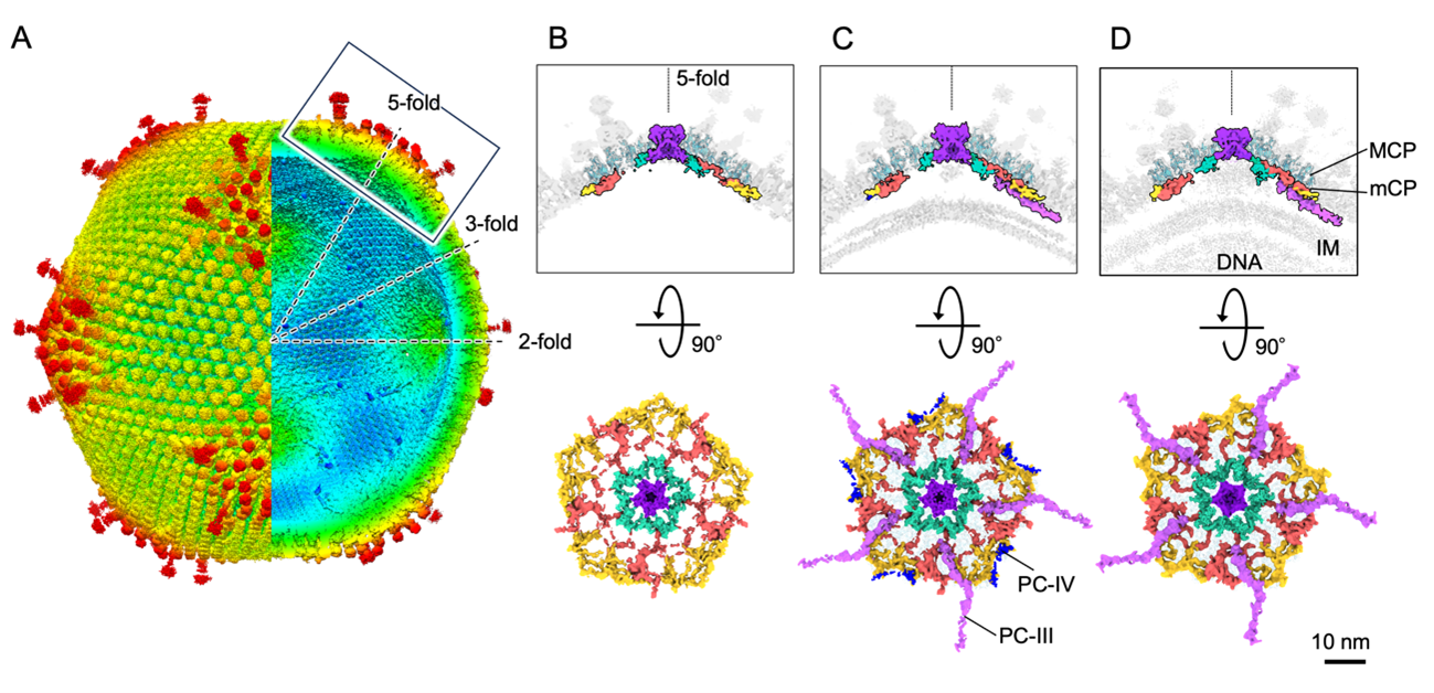
Figure 1. Medusavirus capsid structure at 7 Å resolution (A: external structure on the left, internal structure on the right) and changes in capsid structure near the 5-fold axis of symmetry (box in A) during the virus particle formation process: capsid only (B), with nuclear membrane (IM) (C), and with nuclear membrane and DNA (D).
Collaborative Researcher
Masaharu Takemura (Tokyo Univ Science)Ryoto Watanabe, Song Chihong, Kazuyoshi Murata (NIPS/ExCELLS/ SOKENDAI)
Funding
This study was supported by KAKENHI (JP17H05825, JP19H04845, JP20H03078), AMED BIDNS (JP18am0101072), NIPS Joint Research (20-239), ExCELLS Joint Research (20-004).Journal article
Title: Subnanometer structure of medusavirus capsid during maturation using cryo-electron microscopyAuthors: Ryoto Watanabe, Chihong Song, Masaharu Takemura, Kazuyoshi Murata
Journal: Journal of Virology
Issue: 98(9), e0043624
Date: 28 August, 2024
URL: https://doi.org/10.1128/jvi.00436-24