Recent Research Results
Experience-dependent emergence of fine-scale networks in visual cortex
September 10, 2014
Visual cortical neurons selectively respond to particular features of visual stimuli and this selective responsiveness emerges from specific connectivity in the cortex. While most visual response properties are basically established by eye opening, they are thereafter modified or refined by visual experience based on activity-dependent synaptic modifications during an early postnatal period. Visual deprivation during this period impairs development of visual functions such as visual acuity. We previously demonstrated that fine-scale networks composed of a population of interconnected layer 2/3 (L2/3) pyramidal neurons receiving common inputs from adjacent neurons were embedded in a small area in rat visual cortex and suggested that this network could be a functional unit for visual information processing. In this study, we investigated the effects of early visual experience on the development of fine-scale networks and individual synaptic connections in rat visual cortical slices. We used two kinds of deprivation, binocular deprivation and dark rearing, which allowed visual inputs with only diffuse light and no visual input, respectively. The probability and strength of excitatory connections to L2/3 pyramidal cells increased during the 2 weeks after eye opening, and these changes were prevented by dark rearing, but not binocular deprivation. Fine-scale networks were absent just after eye opening and established during the following 2 weeks in rats reared with normal visual experience, but not with either type of deprivation. These results indicate that patterned vision is required for the emergence of the fine-scale network, whereas diffuse light stimulation is sufficient for the maturation of individual synapses.
Ishikawa, A.W., Komatsu, Y., Yoshimura, Y. (2014) Experience-dependent emergence of fine-scale networks in visual cortex. J Neurosci. 34(37):12576-86
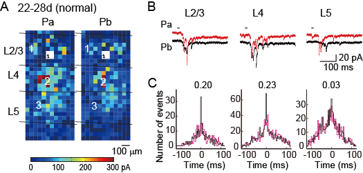
A, The color of each square indicates the sum of the amplitudes of EPSCs evoked in pyramidal cells Pa and Pb by photostimulation at that site in a slice prepared from a normally reared rat at 22-28d.
B, EPSCs evoked in Pa (red) and Pb (black) by photostimulation at representative sites (squares numbered in A) in L2/3, L4 and L5. C, Non-shifted (black) and shifted (magenta) cross-correlograms computed from EPSC data collected upon photostimulation in each layer. The figures attached to the plots indicate the correlation probability values in the cell pair.
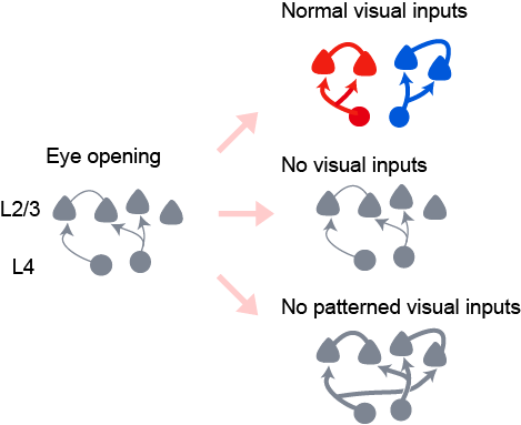
The development of excitatory synaptic connections from L4 cells to L2/3 pyramidal cells during the period from eye opening (left) to 2 weeks after eye opening is shown schematically for rats reared normally (upper right), in darkness (middle right) and with binocular deprivation (lower right). The thickness of lines indicates the strength of synaptic connections.