3.神経情報処理の分子細胞基盤

1.異種シナプス間拡散性クロストーク

  神経情報は、多数のニューロン(neuron:神経細胞)からなる神経回路を伝達される過程で様々な処理(変換・統合)を受けます。 ニューロンとニューロンの間の情報伝達を仲介する構造をシナプス(synapse)と呼び、一般に神経情報は、送信側のニューロンであるシナプス前細胞(presynaptic cell)から、 受信側のシナプス後細胞(postsynaptic cell)へと一方向に伝達されます。シナプスは、シナプス後細胞を興奮させる興奮性シナプスと、 シナプス後細胞を抑制する抑制性シナプスに大別することができます。中枢神経では、主にグルタミン酸(glutamic acid)とγ-アミノ酪酸(g-aminobutyric acid, GABA)が 情報伝達物質として働き、シナプス後細胞の膜興奮性をそれぞれ亢進もしくは抑制します。しかし最近、神経情報を処理していく過程において、 シナプス伝達は一方通行に進む(anterograde transmission)のみならず、逆走したり(retrograde transmission)、脇道にそれたり(heterosynaptic transmission) することが分かってきました。

  脇道にそれる例として私たちは、脳幹の下オリーブ核から小脳プルキンエ細胞(Purkinje cell)に投射する登上線維(climbing fiber)の興奮性伝達物質 (グルタミン酸と推定)が、分子層介在ニューロン(molecular layer interneuron)から同じプルキンエ細胞に入力するGABA作動性のシナプス伝達を抑圧することを見出しました(図1)。 抑制性入力が減弱するため、登上線維入力を受けて興奮したプルキンエ細胞の興奮性は、 さらに強化される[即ち、脱抑制(disinhibition)]と考えられます(Satake et al., 2000)。


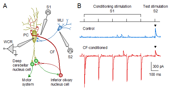
図1:小脳皮質の異種シナプス間拡散性クロストーク。 (A)全細胞パッチクランプ法(whole-cell recording, WCR)により、プルキンエ細胞(PC)からシナプス後電流を記録する。 ガラス電極(S1)を用いて登上線維(CF)に条件刺激を与えた後、別のガラス電極(S2)にて介在ニューロン(MLI)軸索にテスト刺激を付加した。 (B)登上線維の反復刺激(5ヘルツで1秒間)に伴い、介在ニューロン−プルキンエ細胞間のGABA作動性伝達(抑制性シナプス後電流)が減弱した(▼印)。
  薬理学実験ならびに免疫組織染色実験の結果から、 登上線維の反復刺激に伴い大量に放出された興奮性伝達物質は、放出部位から拡散して(脇道にそれて)近傍に存在する介在ニューロン終末の AMPA作動性グルタミン酸受容体[イオン透過型受容体(ionotropic receptor)の一種]を活性化することにより、 介在ニューロンのGABA放出を減弱させた[シナプス前抑制(presynaptic inhibition)]と考えています(図2)(Satake et al., 2004, 2006)。

  また、ニューロン型グルタミン酸輸送体EAAT4(プルキンエ細胞特異的に発現)とグリア型グルタミン酸輸送体GLAST[シナプスを被覆するバーグマングリア(Bergmann glia)に発現]は、 グルタミン酸が登上線維終末から籠細胞終末に拡散していく過程をそれぞれ独立に制御できることを明らかにしました(Satake et al., 2010)。プルキンエ細胞は小脳皮質で唯一の出力細胞です。 また、登上線維−プルキンエ細胞間の興奮性シナプスは極めて強固なため、世界中で古くから研究されてきた重要なシナプスです。この重要なシナプスが、同時に異種シナプス抑制を 起こすことにより小脳出力を強化するという巧妙な仕掛けを併せ持つことの意義を考えると、 (伝達物質が)脇道にそれる現象を研究することも、(脳の情報処理機構を理解するには)あながち脇道にそれた研究ではないのではないかと思えてきます。
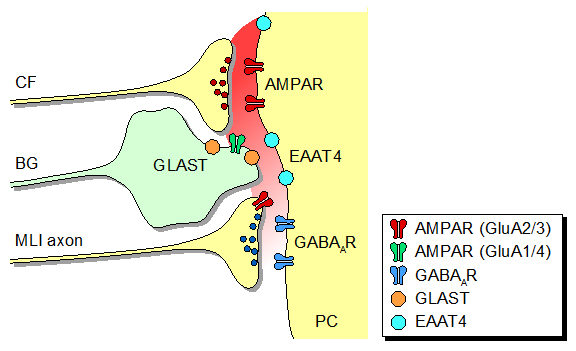
図2:登上線維(CF)から放出された興奮性伝達物質は、 後シナプス性AMPA受容体を活性化してプルキンエ細胞を興奮させるとともに、シナプス間隙の外にまで拡散する。 拡散した伝達物質は、介在ニューロン(MLI)軸索終末の前シナプス性AMPA受容体(GluA2/3)に作用することにより、 介在ニューロンのGABA放出を抑制する。こうしたシナプス外拡散は、プルキンエ細胞やバーグマングリア(BG)の グルタミン酸輸送体(EAAT4/GLAST/GLT-1)によって常に阻害されているものの、反復刺激に伴い大量に放出された登上線維伝達物質は、 輸送体の回収能を超えて介在ニューロンのAMPA受容体に到達する。

2.シナプス小胞の多重性放出

    中枢神経の軸索終末では、活動電位の伝播に伴い、1個のシナプス小胞が開口放出されると考えられてきました[単一性放出(univesicular release)]。 その一方で、1回の活動電位当たり、複数のシナプス小胞が複数の放出部位から同期的に、もしくは単一の放出部位から連続的に放出される、多重性放出(multivesicular release)の存在も指摘されています。シナプス小胞の単一性/多重性放出の違いは、神経終末のCa2+動態や放出機構の性質の違いによって生じると推定されてきました。 しかし、神経終末のシナプス小胞放出ダイナミクスを制御するメカニズムは、まだほとんど解明されていません。

  私たちは、ラット小脳顆粒細胞(granule cell)から分子層介在ニューロンへの興奮性シナプスにおいて、顆粒細胞の2回パルス刺激(30〜100ミリ秒間隔)に伴い、 介在ニューロンから記録される興奮性シナプス後電流(excitatorypostsynaptic current, EPSC)の2回目の振幅値と減衰時定数(t)が、 刺激間隔に依存して一過性に増大する現象を発見しました(図3)。減衰時間のペアパルス増大は、シナプス小胞の放出多重性に伴う 興奮性神経伝達物質グルタミン酸のシナプス間蓄積によって引き起こされたと推定しています(Satake et al., 2012)。


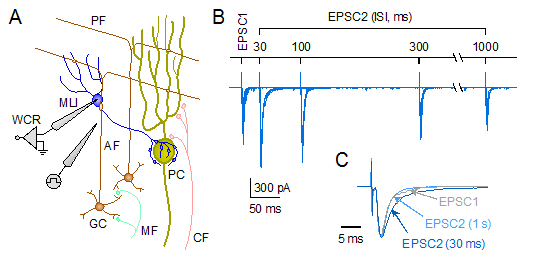
図3:小脳顆粒細胞−介在ニューロン間興奮性シナプスにおけるペアパルス増強。 (A)顆粒細胞(GC)軸索(上行性線維:ascending fiber, AF)を電気刺激し、介在ニューロン(MLI)からEPSCを記録する。 (B)2回のパルス刺激に伴い、2回目EPSC(EPSC2)の振幅値と減衰時定数(t)が一過性に1回目EPSC(EPSC1)より増大する。苔状線維mossy fiber, MF、平行線維parallel fiber, PF。(C)振幅を揃えたEPSCトレース[灰色:EPSC1、濃青色:EPSC2(ISI = 30 ms)、薄青色:EPSC2(ISI = 1 s)]。


  しかし、EPSCの減衰時間が多重性放出によって決定されるという現象はこれまで報告がなく、 その分子的背景も明らかでありませんでした。そこで、EPSC減衰時間のペアパルス増大を実験モデルとして、 多重性放出の発現メカニズムを追究しました。その結果、顆粒細胞の多重性放出は、 (1)複数のCav2.1チャネルを介して顆粒細胞の軸索終末に流入したCa2+が流入部位から周囲に拡散して蓄積すること、 (2)蓄積したCa2+がチャネル近傍に局在するシナプス小胞/Ca2+センサー複合体のみならず、 (3)遠位の小胞/Ca2+センサー複合体にも作用して複数のシナプス小胞を放出させることによって惹起されたことなどが明らかになりました (Satake and Imoto., 2014)。Ca2+チャネルサブタイプに依存した異なるシナプス小胞放出過程(単一性放出と多重性放出)の発見は、 脳の情報処理プロセスに多様性を生み出すメカニズムの一つとして、今後の神経回路研究に新しい道を拓くと期待されます(Satake et al., 2015)。


参考文献

Satake S., Saitow F., Yamada J.,Konishi S. (2000) Synaptic activation of AMPA receptors inhibits GABA release from cerebellar interneurons. Nat. Neurosci. 3, 551-558. [PubMed]

Satake S., Saitow F., Rusakov D.,Konishi S. (2004) AMPA receptor-mediated presynaptic inhibition at cerebellar GABAergic synapses: a characterization of molecular mechanisms. Eur. J. Neurosci. 19, 2464-2474. [PubMed]

Satake S., Song S.-Y., Cao Q., Satoh H., Rusakov D. A., Yanagawa Y., Ling E.-A., Imoto K.,Konishi S. (2006) Characterization of AMPA receptors targeted by the climbing fiber transmitter mediating presynaptic inhibition of GABAergic transmission at cerebellar interneuron-Purkinje cell synapses. J. Neurosci. 26, 2278-2289. [PubMed]

Satake S., Song S.-Y., Konishi S.,Imoto K. (2010) Glutamate transporter EAAT4 in Purkinje cells controls intersynaptic diffusion of climbing fiber transmitter mediating inhibition of GABA release from interneurons. Eur. J. Neurosci. 32, 1843-1853. [PubMed]

Satake S., Inoue T.,Imoto K. (2012) Paired-pulse facilitation of multivesicular release and intersynaptic spillover of glutamate at rat cerebellar granule cell-interneurone synapses.  J. Physiol. 590, 5653-5675. [PubMed]

Satake S., Imoto K. (2014) Cav2.1 channels control multivesicular release by relying on their distance from exocytotic Ca2+ sensors at rat cerebellar granule cells. J. Neurosci. 34, 1462-1474. [PubMed]

Satake S., Inoue T., Imoto K. (2015) Synaptic multivesicular release in the cerebellar cortex: its mechanism and role in neural encoding and processing. Cerebellum in press. [PubMed]
----------------------------------------

もとに戻る


3.Molecular and cellular mechanisms underlying neural encoding and processing

1.Transmitter diffusion-mediated crosstalk between heterologous neurons

At most central excitatory synapses, the acidic amino acid L-glutamate has been believed to serve as a major neurotransmitter and mediate fast excitatory neurotransmission through activation of postsynaptic ionotropic glutamate receptors. Considerable evidence is accumulating that glutamate can also act on presynaptic receptors to mediate a variety of modulatory actions on neurotransmitter release by activating either autoreceptors within single synaptic connections and heteroreceptors in adjacent distinct synapses.

Previously, we reported that the excitatory transmitter of the climbing fibers (CFs) originated from the inferior olivary nucleus to the cerebellar cortex could elicit not only a powerful excitation in postsynaptic Purkinje cells (PCs) but also an suppressive modulatory action on GABA (g-aminobutyric acid) release from molecular-layer interneurons (MLIs) converging onto the same PCs (Satake et al., 2000) (Fig. 1). Both postsynaptic excitation and presynaptic inhibition were likely mediated by activation of AMPA-sensitive glutamate receptors (AMPARs) (Satake et al., 2004).

Fig. 1: Heterosynaptic crosstalk in rat cerebellar cortex. (A) Diagram showing neural elements, and electrodes for recording and stimulation. Whole-cell recording, WCR. (B) Repetitive activation of the CF (by S1 electrode at 5 Hz for 1 s) inhibited GABAergic transmission at MLI-PC synapses (evoked by S2 electrode, arrowhead).

A line of evidence from pharmacological and morphological experiments suggested that the CF transmitter released upon repeated synaptic activation escapes local glutamate uptake and extrasynaptically diffuses from the release sites to reach remote AMPARs expressed on the axon terminals of neighboring MLIs (Satake et al., 2006). We more recently reported that glutamate transporters (EAAT4 and GLAST) play unique roles in determining the degree of CF-induced suppression of cerebellar GABAergic transmission by influencing the glutamate concentration in the route of its extrasynaptic diffusion (Satake et al., 2010) (Fig. 2).
Fig. 2: CF transmitters spill out of the synaptic cleft and activate AMPARs expressed in the MLI terminals (GluA2/3 subtypes), leading to inhibition of GABA release from the MLI. Extrasynaptic concentration of the CF glutamate is controlled by excitatory amino acid transporters (EAATs) expressed in postsynaptic target PCs (EAAT4 subtype) and perisynaptic processes of Bergmann glia (BG; GLAST subtype).

2.Synaptic multivesicular release

The number of vesicles released for fast neurotransmission plays a major role in determining the strength of the postsynaptic response. The quantal output of a single axon terminal is generally restricted to one vesicle per presynaptic action potential (univesicular release), while the concomitant release of multiple vesicles per action potential (multivesicular release, MVR) has been reported in some CNS synapses. However, the mechanisms responsible for MVR remain unsolved. Paired-pulse facilitation (PPF) is a ubiquitous form of presynaptic short-term plasticity. We recently reported that repetitive activation of rat cerebellar granule cell (GC) axons at short intervals (30?100 ms) causes both facilitation of the peak amplitude (PPFamp) of the second EPSC (EPSC2) recorded from MLIs and prolongation of the EPSC2 decay time (PPPdecay) relative to those of the first EPSC (EPSC1) (Fig. 3). PPFamp and PPPdecay are elicited by different mechanisms. PPFamp results from transient increases in release probability and MVR. On the other hand, PPPdecay is elicited by extrasynaptic spillover of MVR glutamate and subsequent glutamate pooling among adjacent active synapses (Satake et al., 2012).


Fig. 3: Different types of paired-pulse modulation at rat cerebellar GC-MLI synapses. (A) Paired-pulse activation of GC ascending fibers (AFs) causes both PPFamp and PPPdecay at GC-MLI synapses. Mossy fiber, MF; parallel fiber, PF. (B) Representative EPSC traces showing PPFamp and PPPdecay. (C) Averaged EPSC1 (light gray trace) and EPSC2 (30-ms ISI, blue trace; 1-s ISI, light blue trace) traces were scaled to the same peak amplitude and superimposed.


Synaptic vesicle release is triggered by Ca2+ influx through several subtypes of voltage-gated Ca2+ channels (VGCCs) located on the presynaptic membrane adjacent to vesicle release sites. In the mammalian CNS, Cav2.1 (P/Q-type) and Cav2.2 (N-type) channels are the dominant VGCC subtypes triggering vesicular neurotransmitter release. More recent empirical evidence suggests that (1) Cav2.1 and Cav2.2 channels differently regulate PPPdecay and PPFamp and (2) the distance between Cav2.1 channels and exocytotic Ca2+ sensors plays a critical role in eliciting MVR (Satake and Imoto, 2014). By transducing presynaptic action potential firings into unique Ca2+ signals and vesicle release profiles, Cav2 channel subtypes and MVR will contribute additional complexity to neural encoding and processing in the cerebellar cortex (Satake et al., 2015).


References

Satake S., Saitow F., Yamada J.,Konishi S. (2000) Synaptic activation of AMPA receptors inhibits GABA release from cerebellar interneurons. Nat. Neurosci. 3, 551-558. [PubMed]

Satake S., Saitow F., Rusakov D.,Konishi S. (2004) AMPA receptor-mediated presynaptic inhibition at cerebellar GABAergic synapses: a characterization of molecular mechanisms. Eur. J. Neurosci. 19, 2464-2474. [PubMed]

Satake S., Song S.-Y., Cao Q., Satoh H., Rusakov D. A., Yanagawa Y., Ling E.-A., Imoto K.,Konishi S. (2006) Characterization of AMPA receptors targeted by the climbing fiber transmitter mediating presynaptic inhibition of GABAergic transmission at cerebellar interneuron-Purkinje cell synapses. J. Neurosci. 26, 2278-2289. [PubMed]

Satake S., Song S.-Y., Konishi S.,Imoto K. (2010) Glutamate transporter EAAT4 in Purkinje cells controls intersynaptic diffusion of climbing fiber transmitter mediating inhibition of GABA release from interneurons. Eur. J. Neurosci. 32, 1843-1853. [PubMed]

Satake S., Inoue T.,Imoto K. (2012) Paired-pulse facilitation of multivesicular release and intersynaptic spillover of glutamate at rat cerebellar granule cell-interneurone synapses.  J. Physiol. 590, 5653-5675. [PubMed]


Satake S., Imoto K. (2014) Cav2.1 channels control multivesicular release by relying on their distance from exocytotic Ca2+ sensors at rat cerebellar granule cells. J. Neurosci. 34, 1462-1474. [PubMed]

Satake S., Inoue T., Imoto K. (2015) Synaptic multivesicular release in the cerebellar cortex: its mechanism and role in neural encoding and processing. Cerebellum in press. [PubMed]

----------------------------------------

 Back