![]()
![]()
![]()
![]()
![]()
![]()

![]() Cells do neither get lean nor grow fat too much.
Cells do neither get lean nor grow fat too much.
There are billions of cells in an adult human body.
Cells continuously take essential substances up from and throw unnecessary substances away to the extracellular fluid during their life.
However, even then, the cell volume is regulated at a constant level. It is due to the volume regulation mechanism that our body
(even only a part of it) does not lose or gain weight in disorder. A variety of proteins located on the cell membrane or
in the cytosol are involved in the cell volume regulation.We are carrying out research works by identifying protein machineries related
to cell volume regulation and by investigating their molecular mechanisms.
![]() Studies about water and ion channels.
Studies about water and ion channels.
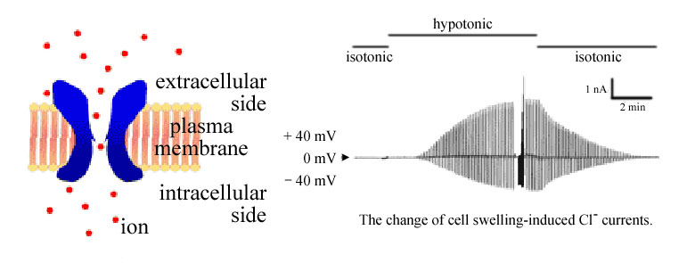
Channels and transporters, which are transmembrane proteins exposed to both extracellular and intracellular fluids,
are essentially involved in the cell volume regulation mechanism. They can carry water, ions, amino acids and so on
and transfer them across the cell membrane. Their activities and characteristics can be, in real time,
observed by measuring the currents produced by charge movements.
Schematic models of cell volume regulation mechanisms.
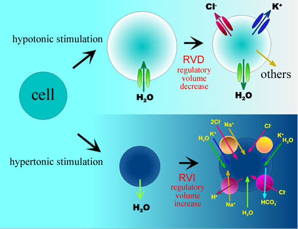
Activation of volume-sensitive Cl- channel upon osmotic cell swelling.
![]() Studies about ATP release from the cell interior.
Studies about ATP release from the cell interior.
Upon osmotic cell swelling, ATP is released from the cytosol to the extracellular fluid. Released ATP is known to upregulate the volume
recovery process after osmotic swelling via stimulation of ATP receptors. However, little is known as to what kind of machinery
(channel, transporter or exocytosis ?) provides the releasing pathway. We are also studying to answer this question.
![]() Dysfunction of cell volume regulation and cell death.
Dysfunction of cell volume regulation and cell death.
Cell death is generally classified into two types. Apoptosis (or programmed cell death) is induced by an innate cell suicide
program installed in genes. Persistent cell shrinkage is one of major hallmarks of apoptosis. On the other hand,
necrosis is an accidental cell death independent of the gene-inherited program. Necrotic cell death is paralleled by persistent cell swelling.
It has recently become clear that the process leading to cell death is closely related to dysfunction of cell volume regulation.
We are investigating the initiation process of cell death from a physiological standpoint.
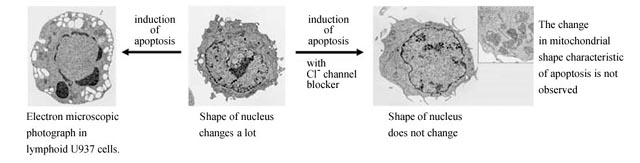
Electron micrographs of changes in cell morphology after apoptotic stimulation in the absence and presence of Cl- channel blocker.
 This site is best viewed with Netscape Navigator 3.0 or later.
We express great thanks to dpi web graphix for the design of our web pages.
This site is best viewed with Netscape Navigator 3.0 or later.
We express great thanks to dpi web graphix for the design of our web pages.
Laboratory of Correlative Physiology