Hypertension is a major risk factor of various diseases including stroke, heart failure, vascular disease, and kidney disease. Angiotensin II, which is produced by the renin-angiotensin system, primarily functions as a physiological regulator of blood pressure and cardiovascular homeostasis, but it also plays a major role in the pathogenesis of hypertension. In the aorta, angiotensin II promotes hypertrophy of vascular smooth muscle cells through angiotensin type 1 receptor (AT1R), resulting in hypertension by increasing arterial wall thickness and vascular resistance. Additionally, angiotensin II induces physiological proliferation of neonatal but not adult vascular smooth muscle cells. How vascular smooth muscle cells determine the responsiveness to angiotensin II under different developmental conditions is mostly unclear.
We found that the purinergic P2Y6 receptor (P2Y6R) promoted angiotensin II-induced hypertension in mice. Mice with a deletion of P2Y6R attenuated an angiotensin II-induced increase in blood pressure and medial hypertrophy (Fig. 1). AT1R and P2Y6R formed stable heterodimers. P2Y6R antagonist MRS2578 disrupted heterodimer formation of AT1R and P2Y6R, and suppressed angiotensin II-induced hypertension in mice.
Furthermore, P2Y6R abundance was developmentally increased in vascular smooth muscle cells. The increased abundance of P2Y6R converted AT1R-stimulated signaling in vascular smooth muscle cells from proliferation to hypertrophy (Fig. 2). These results suggest that increased formation of AT1R-P2Y6R heterodimers with age may increase the likelihood of hypertension induced by angiotensin II.
This work was done by collaboration with Caroline Sunggip from Universiti Malaysia Sabah, Hidetoshi Tozaki-Saitoh, Tomomi ide, Hitoshi Kurose, Makoto Tsuda and Kazuhide Inoue from Kyushu University, Katsuya Hirano from Kagawa University, and Jean-Marie Boeynaems and Bernard Robaye from Université Libre de Bruxelles. This research was supported by grants from the Japan Science and Technology Agency, the Precursory Research for Embryonic Science and Technology Program, Grants-in-Aid for Scientific Research, and Platform for Drug Discovery, Informatics, and Structural Life Science from the Ministry of Education, Culture, Sports, Science and Technology, Japan; Kimura Memorial Heart Foundation, Daiko Foundation and NOVARTIS Foundation (Japan) for the Promotion of Science.
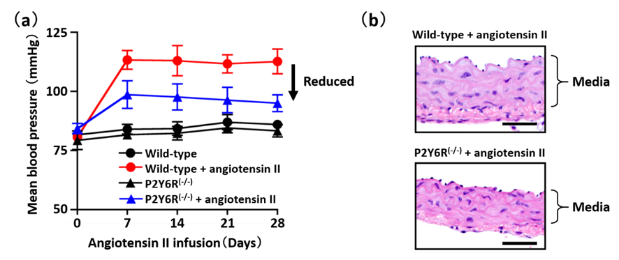
Fig. 1. P2Y6R deletion attenuates angiotensin II-induced hypertension. (a) Mean blood pressure of wild-type or P2Y6R(-/-) mice treated with or without angiotensin II. (b) Hematoxylin and eosin-stained images of aortae from wild-type or P2Y6R(-/-) mice treated with angiotensin II.

Fig. 2. Developmental increase in P2Y6R abundance enhances angiotensin II-induced hypertrophic response. P2Y6R abundance was developmentally increased in vascular smooth muscle cells. In neonatal cells, angiotensin II induced proliferation through AT1R monomer, whereas angiotensin II induced hypertrophy of adult vascular smooth muscle cells through AT1R-P2Y6R heterodimer.


