As a mission to be the inter-university research institute, NIPS conducts joint studies with researchers from domestic or foreign universities and other research institutes. NIPS provides specialized equipment, large-scale equipment, and research facilities, and develops new equipment for morphological and functional 4D imagings of various organs such as the brain.
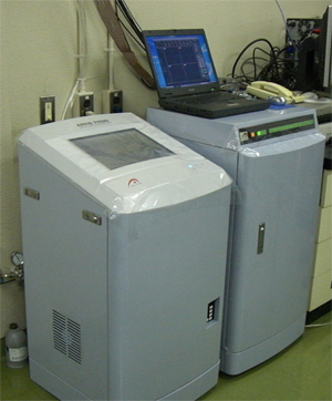
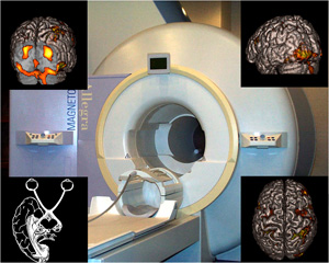 MRI is an imaging technique that utilizes the nuclear magnetic resonance of hydrogen and other atoms. MRI enables not only detailed structural imaging of the brain but also the localization of brain function by measuring regional cerebral blood flow (functional MRI). The NIPS installed an ultra-high field (7T) MRI system (Siemens Magnetom 7T) in 2014. In fiscal year 2023, we also installed a 3T MRI system with strong gradient (Siemens Magnetom Cima.X). Furthermore, the NIPS installed an 11.7T Bruker preclinical MRI system in fiscal year 2025.
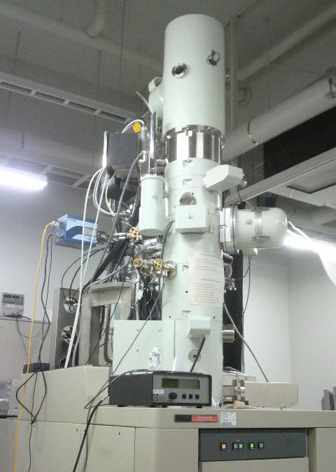 Electron cryomicroscope is an electron microscope developed for observing close-to-life state biological samples with a combination of rapid freezing and ice embedding sample preparation methods. Biological specimens up to 200 nm thicknesses can be observed with high-resolution and high-contrast. Ultrastructure analyses of protein molecules, viruses, bacteria, cultured cells, and frozen tissue sections are performed with this novel microscopic system.
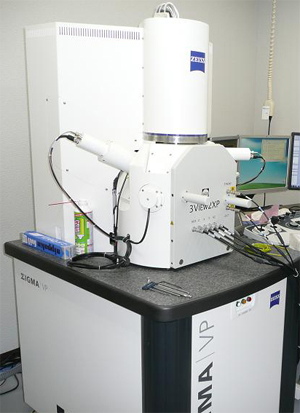 Serial block-face scanning electron microscope(SBF-SEM) is an advanced 3-D nano-imaging equipment. Two different types of SBF-SEM are available; high-resolution and wide-area types. Resin-embedded biological specimens are sliced by a diamond knife equipped inside the chamber, and the block-face images are acquired by scanning electron microscopy (SEM). 3-D structures of the specimens are finally reconstructed from the acquired serial block-face images. 3-D structures of large biological specimens like brain tissue can be visualized at the resolution of several nanometers.
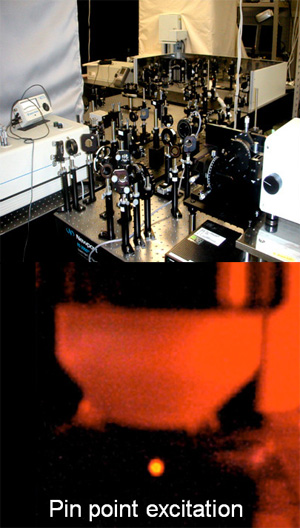 Multi-photon excitation is a method to visualize living tissue by exciting the fluorescence molecules with the tightly focused near-infrared femtosecond pulse laser. Since the longer wavelength is used for multi-photon excitation, it has a superior deeper tissue penetration and reduced phototoxicity compared with single-photon excitation. Our 2-photon microscopes have the top-level specification for deep tissue imaging and can be applied to the imaging of neurons and glial cells in deep tissues such as the mouse brain. Recently, we also developed a 2-photon fluorescence imaging microscope that can be applied to image protein-protein interaction and the protein activity.
 We analyze the following physiological parameters in mice:
(A) Evaluation of behaviors related to emotions, learning, and memories, and analyses of neural and muscular activities,
(B) Non-invasive 4D cardiac function and capillary blood flow ultrasound imaging in mice,
(C) Functional analysis of neuroimmune interactions in mouse models of diseases, (D) Multicellular activity measurement and manipulation in vivo,
(E) Physiological measurements and analysis in vivo
[Major apparatuses]
Brain wave–measuring apparatus, Electromyograph, Telemetry automatic measurement system for chronic experiments, 4D ultrasound imaging device VEVO3100, Isolated heart perfusion system, Open field test analyzer, Light/dark transition test device, Barnes circular maze test device, Elevated plus-maze test analyzer, Forced swimming test analyzer, Rota-rod test analyzer, Passive avoidance test analyzer, Fear conditioning test analyzer, Morris water maze pool, IntelliCage: group-housed automated high-throughput behavioral and cognitive screening system, Nikon A1MP+holographic microscope, The head-mounted miniature microscope, X-ray irradiation device, silicon CMOS digital neural probe.


