Researchers Develop a Carbon Nanotube Tape for Facilitating Serial-Section Electron Microscopy used for Reconstructing Brain Circuits.
Scientists from Japan’s National Institute for Physiological Sciences and their collaborators have made a substantial technological development for automatically collecting thin brain sections that are then electron microscopically imaged and used for mapping the brain.
Okazaki, Japan – What is the neural basis of the mind? The answer may be found in the precise "wiring diagram" of our brains. The “connectome,” the map of synaptic connections between all neurons, (Figure 1) indicate exactly where signal transmission occurs. Little is known, however, about the details of this wiring diagram because most existing methods for mapping the brain's circuits lack the resolution required to image the brain's tiniest processes, which can be less than 50 nm in diameter. These wiring diagrams not only help us understand normal brain function but can also be used to diagnose neurodegenerative diseases such as schizophrenia, Alzheimer's, and autism. Electron microscopy is one of the few methods that has the required resolving power to image these fine neural processes.
In order to fully grasp the microstructure of the brain, an ultra-thin section of about 50 nm (approximately 1/2000 the width of a human hair) is cut with a microtome and imaged with an electron microscope. By using an automatic ultrathin-section collecting machine called an ATUM (Glossary term #1) to collect consecutive sections for imaging, laborious manual efforts are already reduced.
However, the team started observing shortcomings when they used existing tapes used for collecting these thin sections. The biggest problem is how the sections accumulate an electrical charge when bombarded with electrons during imaging. This prevents good imaging of neural ultrastructure because electrons that are trapped on the sample can repel incoming electrons, causing them to be displaced. Conductive tape allows the electrons in the section to escape through grounding, preventing these imaging issues. Conventionally, researchers use a plastic tape called "Kapton tape," which can be made conductive by carbon deposition on its surface. However, this carbon-coated Kapton tape is not sufficiently conductive for imaging with high currents, so it’s not optimal for taking high resolution images, where a charge can build up over a small area.
The research team found that, by adding carbon nanotubes (CNT) to coat a conventional plastic tape (PET) (Glossary term #2), conductivity issues were resolved. The inclusion of carbon nanotubes in the tape created a conductive tape that allowed sections to be imaged without further conductive coating and without any image aberrations (Figure 2). They called the combination CNT-PET tape. The advance made with CNT-PET tape will make it even easier to collect and image serial brain sections using the ATUM-SEM pipeline, now with the image quality necessary to identify all synaptic contacts, ultimately leading to the reconstruction of the whole mouse brain wiring diagram.
###
Glossary:
ATUM - Automated Tape-collecting Ultra-Microtomy is essentially a conveyor belt that automatically collects conventionally ultramicrotomed sections onto tape. It was developed by Jeff Lichtman's group at Harvard University in the USA (References). About 50 units are in use around the world, with four in Japan.
CNT-PET - Carbon Nano-Tube-coated Poly-Ethylene Terephthalate. A type of polymer-composed tape that is coated in carbon nanotubes to make it electrically conductive. Carbon nanotubes are an allotrope of carbon composed of a six-membered ring structure and consists of tubes (cylinders) with a diameter of several nanometers. It is a new material that is also excellent in flexibility with very high conductivity.
Brief summary:
1. As a result of using the tape coated with the carbon nanotubes in electron microscopy, we succeeded in imaging nanoscale circuits in brain tissue.
2. We improved the tissue processing method and optimized it for electron microscopic observation, resulting in clearer images of synapses.
Contribution to Society:
This study contributes to the establishment of new methods for analyzing the structure of the neural circuits of the brain. We expect that this will be an important technology for analyzing abnormalities of the neural circuit structure of the brain from patients suffering from neurological diseases, such as Alzheimer's, autism and schizophrenia.
Carbon nanotube tape for serial-section electron microscopy
Yoshiyuki Kubota, Jaerin Sohn, Sayuri Hatada, Meike Schurr, Jakob Straehle, Anjali Gour, Ralph Neujahr, Takafumi Miki, Shawn Mikula, Yasuo Kawaguchi
The paper will be published in online version of Nature Communications https://www.nature.com/ncomms/
1000 London time (GMT) / 0500 US Eastern Time
1900 Japanese time / 2100 Australian Eastern Time
on 30th January 2018.
Summary Text:
An international research team centered at Japan’s National Institute for Physiological Sciences developed a carbon nanotube tape for facilitating serial-section electron microscopy used for reconstructing brain circuits.
Keyword: Neural circuits
Additional keywords: Electron microscopy, Carbon nanotubes, Ultramicrotomy
Cell. 2016 Mar 24;165(1):192-206. doi: 10.1016/j.cell.2016.02.033.
The Fuzzy Logic of Network Connectivity in Mouse Visual Thalamus.
Morgan JL, Berger DR, Wetzel AW, Lichtman JW.
Cell. 2015 Jul 30;162(3):648-61. doi: 10.1016/j.cell.2015.06.054.
Saturated Reconstruction of a Volume of Neocortex.
Kasthuri N, Hayworth KJ, Berger DR, Schalek RL, Conchello JA, Knowles-Barley S, Lee D, Vázquez-Reina A, Kaynig V, Jones TR, Roberts M, Morgan JL, Tapia JC, Seung HS, Roncal WG, Vogelstein JT, Burns R, Sussman DL, Priebe CE, Pfister H, Lichtman JW.
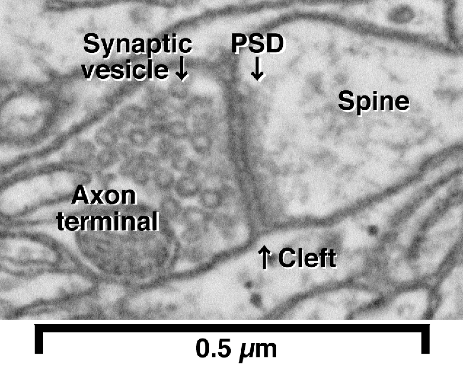
Figure 1. The brain consists of neurons and their connections to each other. Shown is an example of a single connection, called a synapse, between two neurons.
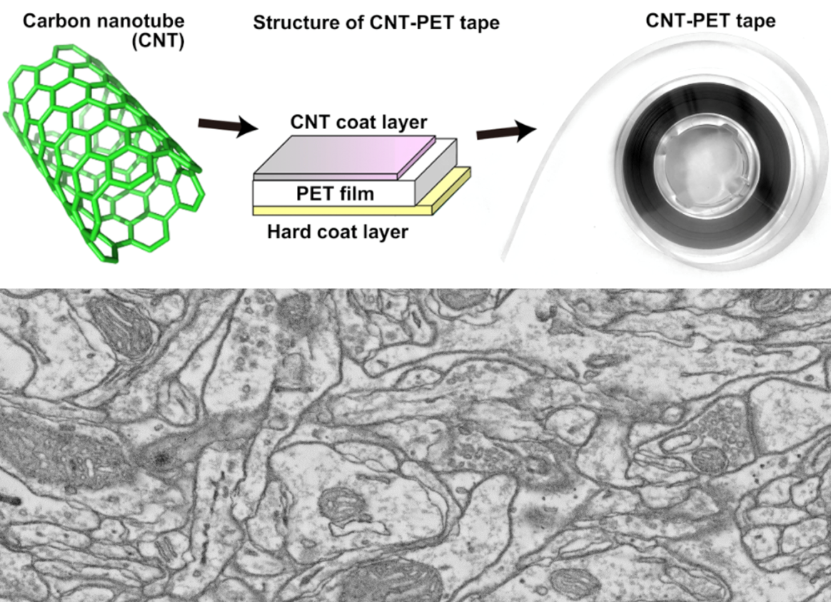
Figure 2. (Upper panel) The new conductive tape is made by coating conventional PET tape with carbon nanotubes (CNTs) to generate CNT-PET tape. (Bottom panel) Sample electron microscopy image from a brain section collected on CNT-PET tape showing good ultrastructure without any charging artefact.
![]()
National Institute for Physiological Sciences (NIPS)


