We are embarking on a new chapter in our pursuit of cutting-edge research in the life and medical sciences. We specialize in applying advanced and innovative imaging techniques to conduct quantitative investigations. Our unique approach involves merging our original, world-class, super-resolution, and ultra-high-speed imaging methods with genetic engineering. This convergence enables us to achieve in vivo visualization of living organisms and to establish a quantitative visual analysis methodology for understanding physiological functions.
Our research also delves into the intricate realm of neural functions, wherein we analyzed neural circuits and activities, biological rhythms, and their molecular foundations. For example, we recently pioneered multiphoton microscopy techniques for deep cross-sectional fluorescence imaging. This microscopy demonstrated neurons within the dentate gyrus of the hippocampus at depths of up to 1.6 mm beneath the brain surface, as well as hippocampal CA1 neuron activity at video rates. Furthermore, our long-term imaging capabilities enable us to study the generation and functions of ultradian and circadian rhythms in living cells. We are also developing super-resolution microscopy, made possible by cutting-edge optical technologies, to explore ultra-micromorphology and molecular dynamics within living cells. Our fast three-dimensional in vivo imaging techniques also offer insights into local neural circuits, endocrine and exocrine glands, and animal and plant models, helping us unravel the fundamental principles governing various physiological functions. Notably, these methods have been instrumental in shedding light on the molecular underpinnings of diseases such as cancer and diabetes.
Our research department actively collaborates with various disciplines, including life sciences, applied physics, material sciences, medicine, and pharmaceuticals. This interdisciplinary synergy enriches our research landscape and allows us to create a vibrant tapestry of knowledge. Our innovative in vivo imaging methodologies are crucial for unlocking the intricate mechanisms governing neural cell physiology, driving us toward new frontiers in scientific understanding.
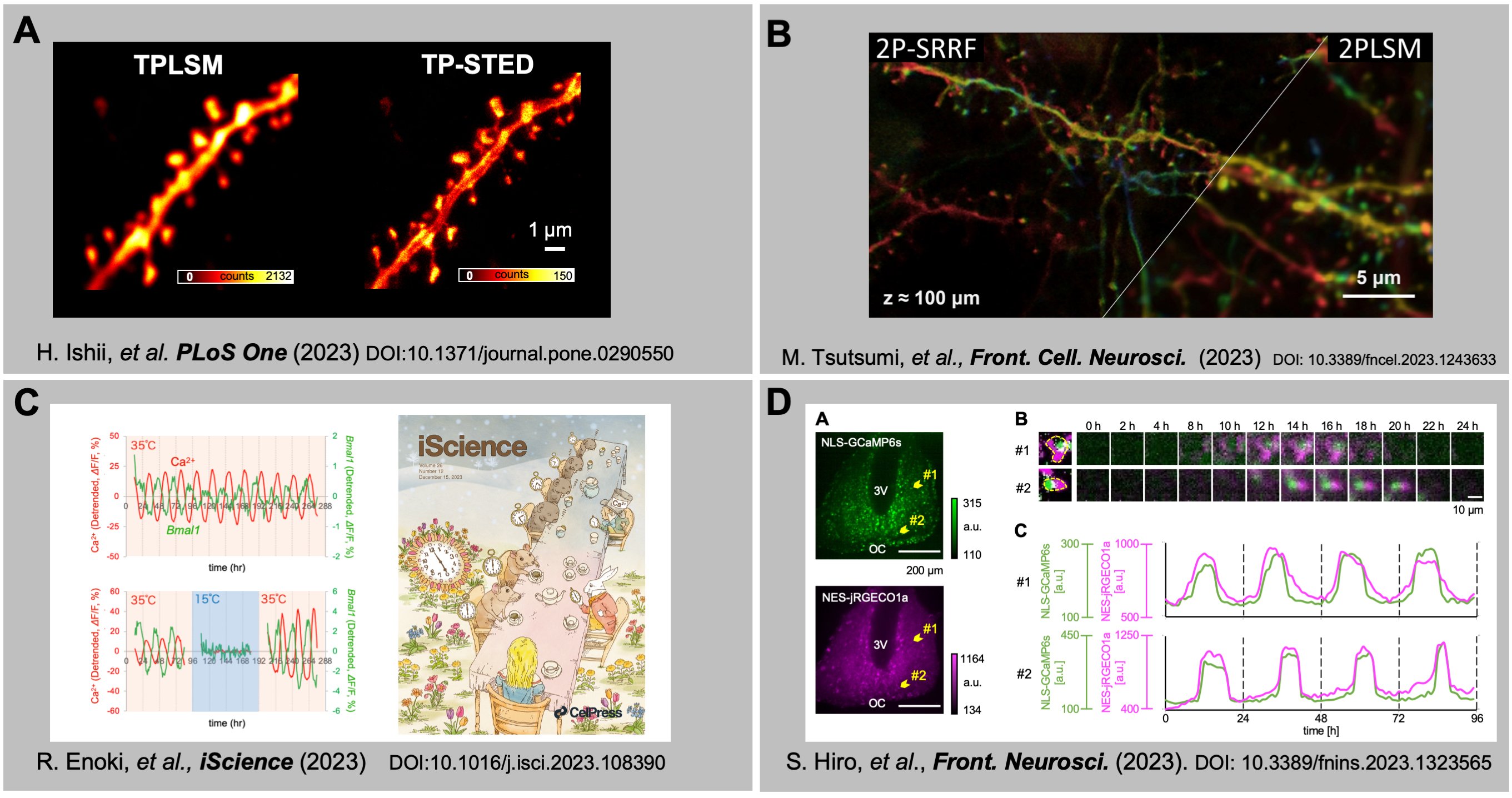
Fig.
(A) Super-resolution imaging with two-photon STED microscopy. (B) Wide-field in vivo imaging using nano-sheets. (C) Cold-induced suspension and resetting of Ca2+ and transcriptional rhythms in the suprachiasmatic nucleus neurons. (D) In-phasic cytosolic-nuclear Ca2+ rhythms in suprachiasmatic nucleus neurons.