Sensory experience during postnatal development is essential for the maturation and refinement of neuronal circuits in the sensory cortex. This process enables the development of cortical functions that are well-suited to the living environment. To elucidate the mechanisms underlying information processing in the sensory cortex and the experience-dependent regulation of this processing, we are studying the relationship between visual functions and the signaling properties of neural circuits, using rat and mouse visual cortex. To this end, we are analyzing the visual responses of cortical neurons using multi-channel electrodes or calcium imaging with two-photon microscopy. Also, we are studying neural circuit properties through a combination of laser scanning photostimulation and whole-cell patch-clamp recording methods in slice preparations, and mapping neural connections morphologically using modern virus tracers. The following is a list of our main ongoing projects:
1.Synaptic plasticity and visual response plasticity in animals at different developmental stages and in animals subjected to manipulations of visual experience during postnatal development.
2.Developmental mechanisms of visual responsiveness, plasticity, and synaptic connections in specific neuron subtypes.
3.Cell-lineage dependent establishment of neuronal connections and visual responsiveness.
We are also conducting collaborative research and seeking graduate students interested in the developmental mechanisms of brain functions.
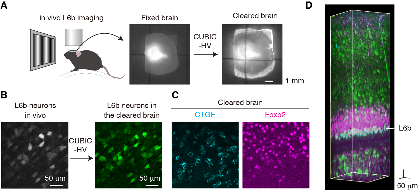
Figure Analysis of visual response plasticity based on cortical neuron subtypes
A) Visual responses were recorded from layer 6b of the primary visual cortex of living mice with two-photon imaging, followed by tissue clearing.
(B) Layer 6b neurons from in vivo two-photon imaging (left) and the same areas from a cleared brain (right).
(C) Most of the recorded L6b neurons expressed CTGF which is a subplate neuron marker. Foxp2 is a marker of cortico-thalamic neurons.
(D) An example of a volumetric image of cleared brain.