Brain processing of the signals ascending through unmyelinated C fibers in humans: an event-related fMRI study
Qiu Y, Noguchi Y, Honda M, Nakata H, Tamura Y, Tanaka S, Sadato N, Wang W, Inui K, Kakigi R: Brain processing of the signals ascending through unmyelinated C fibers in humans: an event-related fMRI study. Cerebral Cortex, 16(9): 1289-1295, 2006.
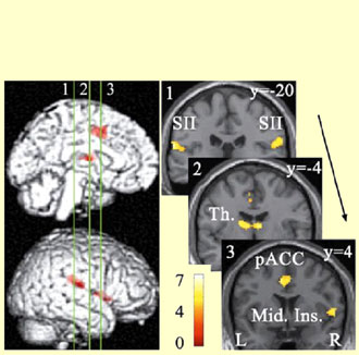
Brain regions commonly activated by C and Ad nociceptor stimulation. Numbered bars in the left panel indicate locations of coronal slices in the right panel. Activated regions overlaid on an anatomically normalized MRI (MNI template) with their corresponding y coordinates (right side). SII = second somatosensory cortex, Th. = thalamus, pACC = posterior portion of the anterior cingulate cortex, Mid. Ins. = middle insula.
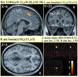
Brain regions differentially activated by C nociceptor stimulation. The activity in these areas was significantly stronger following the stimulation of C nociceptors than Ad nociceptors (P<0.001, uncorrected) and overlaid on an anatomically normalized MRI (MNI template). BA = Brodmann's area, Ant. Ins. = anterior insula, L. = left, R. = right.


