Researchers centered at Japan’s National Institute for Physiological Sciences reveal details of the structure of compacted DNA transiently formed at the time of cell division in cyanobacteria.
Okazaki, Japan –
Eukaryotic cells, including human cells, form paired condensed chromosomes before cell division. The paired chromosomes are then equally divided into the daughter cells. Prokaryotic cells, including bacteria, do not have such a DNA distribution system.
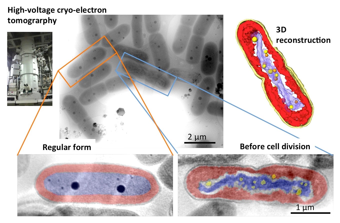
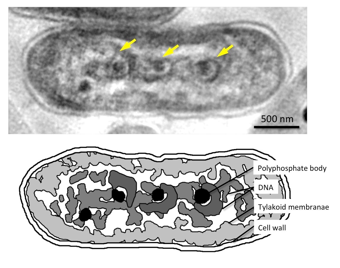
Researchers working at the National Institute for Physiological Sciences (NIPS) and Saitama University have discovered that the photosynthetic cyanobacterium Synechococcus elongatus shows eukaryotic condensed chromosome-like DNA compaction prior to cell division and were able to reveal details of the transiently formed structure. The bacteria divide at the moment of transition from day to night if cultivated under rigorous 12-h light/12-h dark cycles. The ephemeral 3D structure of the compact DNA was captured by high-voltage cryo-electron tomography following rapid freezing. The researchers show that the structure is similar to the condensed chromosomes in eukaryotic cells. The compacted DNA also includes many small and paired polyphosphate bodies, some of which seem to maintain contact with DNA that appears to twist away from them, indicating that they may act as suppliers and regulators of phosphate for DNA synthesis. These new findings may deepen our understanding of how the eukaryotic cell arrived at a system using condensed chromosomes in the long evolution of biological cells.
For the study, published in Scientific Reports, the researchers used a combination of rigorous synchronized cultivation and sophisticated imaging by high-voltage cryo-electron tomography after rapid freezing to depict the transient 3D structure of the compacted DNA in cyanobacteria as thick as 1 μm. The polyphosphate bodies included in the compacted DNA may play a role as phosphate suppliers for DNA synthesis, which are duplicated alongside the DNA duplication.
“We know that the difference between prokaryotes and eukaryotes is in the DNA packaging and delivering system, where eukaryotes have developed a nuclear membrane to protect their genes and form condensed chromosomes to properly parcel the genome into the daughter cell, whereas prokaryotes do not have such a system” corresponding author Kazuyoshi Murata says. “Our new findings throw light on the duplication and segregation mechanisms of cyanobacterial DNA and point to an important role for polyphosphate bodies. They may give hints as to by what steps the evolution from prokaryote to eukaryote took place. ”
The article “Ultrastructure of compacted DNA in cyanobacteria by high-voltage cryo-electron tomography” was published in Scientific Reports at www.nature.com/articles/srep34934 .
Primary Keywords: DNA compaction
Additional Keywords: Cyanobacteria, High-voltage cryo-electron microscope, Microbiology
Twitter Comment: NIPS researchers reveal the ultrastructure of DNA compaction in a cyanobacteria using high-voltage cryo-electron tomography
![]()
![]()
National Institute for Physiological Sciences (NIPS)
Saitama University


