|
The functional roles of the surviving subplate neurons remain largely unknown. Researchers from NIPS in Japan performed two-photon Ca2+ imaging from L6b neurons with post hoc 3D immunohistochemistry of a sublate neuron marker. L6b/ surviving subplate neurons in V1 showed clear responses to visual stimuli. Chronic two-photon imaging showed that L6b neurons exhibited ocular dominance plasticity. These results provide strong evidence that surviving subplate neurons exhibit sensory responses and experience-dependent plasticity. |
The mammalian cerebral cortex consists of six layers, with distinct roles in information processing. At the bottom of the neocortex, on the boundary between the gray matter and white matter, there is a thin sheet of neurons called L6b. L6b neurons are thought to be remnants of subplate neurons that transiently form neural circuits during perinatal development and guide cortical maturation. Most subplate neurons undergo cell death, while some survive. However, the functional properties of L6b/surviving subplate neurons remain largely unknown. Taisuke Yoneda, Kenji Hayashi, and Yumiko Yoshimura at the National Institute for Physiological Sciences have found that L6b/ surviving subplate neurons in the primary visual cortex (V1) exhibit sensory responses and experience-dependent plasticity in juvenile mice. They published their findings in PNAS.
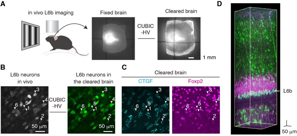
Researchers performed functional measurements of surviving subplate neurons by using two-photon Ca2+ imaging with post hoc tissue clearing and 3D immunohistochemistry of a subplate neuron marker. Most of the recorded L6b neurons expressed a subplate neuron marker and they demonstrated broadly tuned visual response properties (Figure).
Then researchers examined whether L6b neurons exhibit experience-dependent plasticity. To this end, they used ocular dominance plasticity as an experimental model. In this model, if one eye is occluded for several days during the sensitive period in juvenile animals, neurons in V1 lose their response to the deprived eye. By using chronic two-photon imaging of visual responses from the same neurons, they found that L6b neurons exhibited ocular dominance plasticity.
“We characterized the visual response properties and functional plasticity in L6b neurons in V1. The surviving subplate neurons may be involved in the experience-dependent maturation of cortical functions and information processing in the mature cortex” Yoneda says.
(A) A fixed brain before (left) and after (right) tissue clearing. (B) L6b neurons from in vivo two-photon imaging (left) and the same areas from a cleared brain (right). (C) Most of the recorded L6b neurons expressed CTGF which is a subplate neuron marker. Foxp2 is a marker of cortico-thalamic neurons. (D) An example of a volumetric image of cleared brain.
Yoneda T., Hayashi K., and Yoshimura Y., (2023) Experience-dependent functional plasticity and visual response selectivity of surviving subplate neurons in the mouse visual cortex. PNAS
https://doi.org/10.1073/pnas.2217011120559
18
3967,4211
4211,18,3967