|
Researchers have discovered that smooth septate junction proteins regulate intestinal stem cell proliferation by removing aPKC from the apical cortex of differentiating progenitor cells in the Drosophila intestine. This process preserves the Kibra activity and prevents excessive growth-promoting signaling. The findings uncover an unexpected signaling function for cell-cell adhesion proteins in maintaining tissue homeostasis and provide new insights into mechanisms that may be relevant to tissue regeneration and cancer development in higher organisms. |
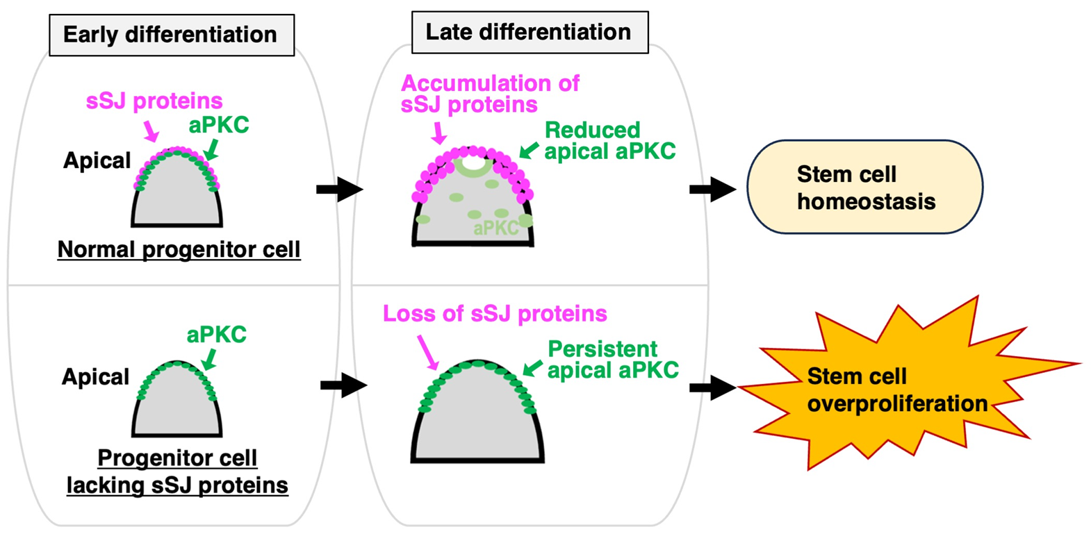 Figure. Loss of sSJ proteins causes persistent apical aPKC in intestinal progenitor cells and promotes stem cell overproliferation
In many animal tissues, stem cells continually divide to create new cells that replace old or damaged ones. However, this capacity requires strict regulation, as uncontrolled stem cell proliferation can disrupt tissue homeostasis and contribute to tumorigenesis.
Researchers at the National Institute for Physiological Sciences have identified a previously unknown mechanism that controls stem cell proliferation in the Drosophila intestine. The study reveals that smooth septate junction (sSJ) proteins, known for their role in cell-cell adhesion, also regulate signaling in differentiating intestinal progenitor cells to prevent excessive stem cell growth.
Unraveling the Adhesion-to-Growth Signal
Previous studies by the research group have shown that the loss of sSJ proteins causes intestinal stem cell overproliferation in fruit flies. However, the molecular mechanism behind this phenomenon remained unclear.
To solve this puzzle, the research team focused on intestinal progenitor cells, which are generated from stem cells and differentiate into mature epithelial cells.
By tracking normal differentiation, the team discovered that sSJ proteins accumulate at the apical region of progenitor cells. Simultaneously, an enzyme named aPKC—a known driver of cell proliferation— is progressively removed from the same region (Fig. top). In contrast, progenitor cells lacking sSJ proteins retain aPKC at their apical region (Fig. bottom).
To determine whether this abnormal localization affects stem cell behavior, the researchers artificially maintained aPKC at the apical region of progenitor cells. This manipulation was sufficient to induce excessive intestinal stem cell proliferation, demonstrating that spatial regulation of aPKC is essential for maintaining normal tissue homeostasis.
Further analyses revealed that persistent apical aPKC suppresses the activity of Kibra, a key activator of the Hippo signaling pathway, which normally restricts cell proliferation. By excluding aPKC from the apical cortex, sSJ proteins preserve Kibra function and prevent the activation of growth-promoting signals.
"Our study demonstrates that differentiating progenitor cells actively regulate stem cell proliferation through cell-cell junction proteins," said Associate Professor Yasushi Izumi. "This finding reveals an unexpected signaling role for cell-cell adhesion molecules beyond simply connecting the neighboring cells. Although our work was conducted in the fruit fly, the underlying principles may contribute to a better understanding of tissue maintenance and cancer development in higher organisms, including humans."
The findings provide new insight into how epithelial tissues coordinate cell differentiation and stem cell activity to maintain tissue integrity and may contribute to future studies on tissue regeneration and cancer biology.
563