Summary Text:
Researchers have developed a new computational method that enables cryo-electron microscopy (cryo-EM) datasets to surpass traditional physical resolution limits after data collection. The technique, called Post-Acquisition Super Resolution (PASR), enhances standard cryo-EM movies through a lightweight pre-processing step applied before motion correction. PASR allows higher-resolution structural information to be recovered from existing datasets without recollecting data at higher magnification and can also be applied to data acquired as super-resolution. The method was successfully tested on multiple datasets, including apoferritin, adeno-associated virus, jack bean urease, and the giant virus Melbournevirus, and proved compatible with standard cryo-EM software pipelines such as RELION and CryoSPARC. PASR may reduce microscope time, storage demands, and data-collection costs while improving structural analysis of large, flexible, or heterogeneous biological complexes.
Full Text:
Cryo-electron microscopy (cryo-EM) has become a powerful tool for determining the structures of proteins, viruses, and molecular complexes at near-atomic resolution. However, the achievable resolution is fundamentally limited by the Nyquist sampling frequency, which is determined by detector pixel size and microscope magnification. Once this physical limit is reached, researchers typically must recollect data at higher magnification, requiring additional microscope time, increased storage capacity, and often fewer particles per image.
Researchers from the Exploratory Research Center on Life and Living Systems and the National Institute for Physiological Sciences have developed a new computational method called Post-Acquisition Super Resolution (PASR), which enables cryo-EM datasets to surpass conventional physical Nyquist limits after data collection. PASR works by computationally subdividing detector pixels before motion correction, allowing subtle particle motion between movie frames to recover higher-frequency structural information.
The method was successfully tested on multiple datasets, including apoferritin, adeno-associated virus, jack bean urease, and the giant virus Melbournevirus, using standard cryo-EM processing software such as RELION and CryoSPARC. PASR improved map quality and enabled higher-resolution reconstructions without visible artifacts. The approach may reduce microscope usage time and data-storage demands while improving analysis of large, flexible, or heterogeneous biological complexes.
Digital Object Identifier (DOI) : 10.1107/S2052252526005348
Related Journal Name: IUCrJ
Article Title: Post-acquisition super resolution for cryo-electron microscopy
Article Publication Date: 2 September 2026
Method of Research: Experimental study
Keywords:
Cryo electron microscopy
Image processing
Expert Contact: Name: Raymond N. Burton-Smith, PhD
Organization: ExCELLS/NIPS
Email: ray@nips.ac.jp
Funder:
1. AMED BINDS under Grant Number JP22ama121005
2. JSPS KAKENHI Grant Number JP22H04926
3. NIPS grant of data science 2022, 2023, 2024
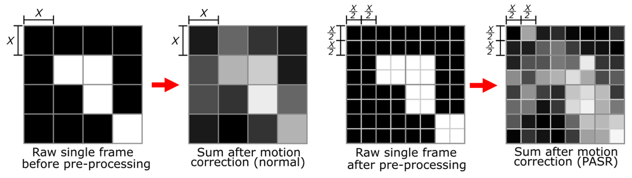 Figure 1: Schematic representations of the PASR concept.
 Comparing to standard motion correction (left panel), sub-dividing pixels prior to motion correction allows for correction of more subtle motion within the captured frames, enabling higher resolution processing. This overcomes physical limitations imposed during acquisition in restrictive situations (right panel).
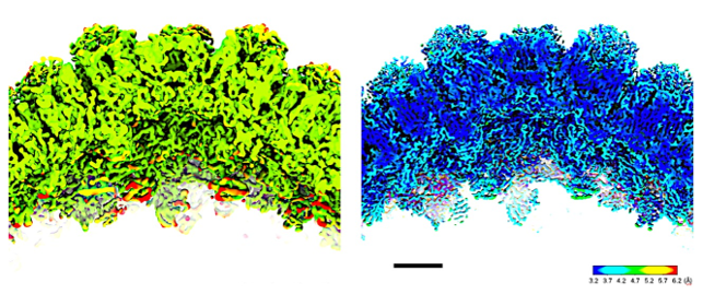 Figure 2: Views of Melbournevirus capsid before (left) and after (right) PASR processing.
 When limited by original acquisition parameters, the fine structure of the capsid is difficult to visualise. After PASR processing, important biological features such as the classic beta-sheet “double jelly roll” fold becomes distinguishable. Scale bar equals 5 nm. Colour coding indicates local resolution, which scales are indicated.
Release Source


The Exploratory Research Center on Life and Living Systems (ExCELLS)
National Institute for Physiological Sciences (NIPS)
5116