Section of Electron Microscopy is a common experimental facility for NIPS and NIBB. Various types of electron microscopes, equipment for sample preparation, and devices necessary for processing digital data acquired by electron microscopy are available in Section Electron Microscopy, enabling a series of work processes of electron microscopy to be carried out. Ultrastructures of tissues, cells and macromolecules are observed using transmission or scanning electron microscopes (JEOL JEM1010, Hitachi HT-7700, Zeiss Σ IGMA). The facility also provides instruments for their specimen preparations, i.e. ultra-microtome (Leica UC7), high-pressure freezing device (BAL-TEC HPM010), freeze fracture and replica machine (BAL-TEC BAF060), vacuum evaporator (JEOL JEE-420), ion coater (JEC-3000FC), etc. Since 2013, Serial block-face scanning electron microscopy (SBF-SEM; Gatan 3view/Zeiss Σ IGMA/VP & MARLIN) and Array tomography SEM system (Zeiss ATLAS5) have been operated to reveal 3D structures of biological thick specimens. The three-dimensional reconstitution of cellular ultra-structures is performed using image analysis software. In particular, the SBF-SEMs are used for many collaborative projects.
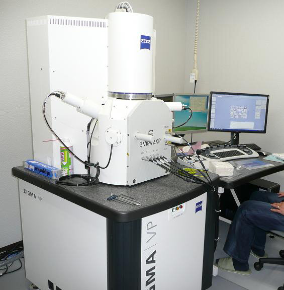
Fig. 1 Serial block-face SEM( SBF-SEM) Gatan 3view - Zeiss Σ IGMA/VP
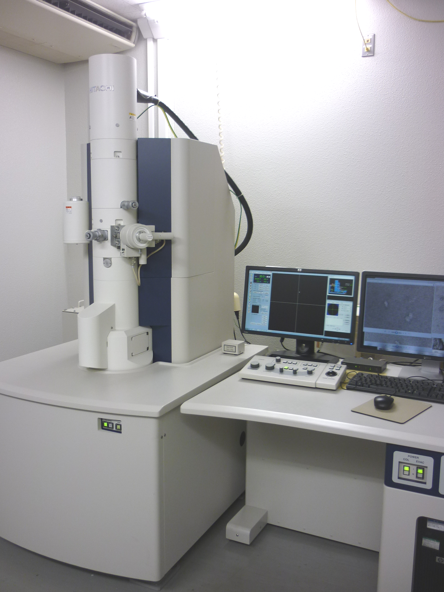
Fig. 2 Transmission electron microscope (TEM) Hitachi HT-7700 equipped with 2kx2k CCD camera


