Our goal is to understand structural changes in biological phenomena including development, functional maintenance and pathophysiology of the nervous system, and elucidate their molecular mechanisms and roles. We utilize various imaging approaches including 3D ultrastructural analyses with serial block-face scanning electron microscopy (SBEM, SBF-SEM) and animal models, and also engage in development of new technologies and many collaborative projects.
We are interested in intercellular associations of the nervous system. Among them, we would like to clarify the structural and functional changes and their molecular background in myelination and myelin diseases. One of our focuses is on mitochondrial dynamics, which are involved in pathophysiology of various diseases. We are trying to clarify the association of mitochondria and myelin diseases, and develop approaches for their regulation.
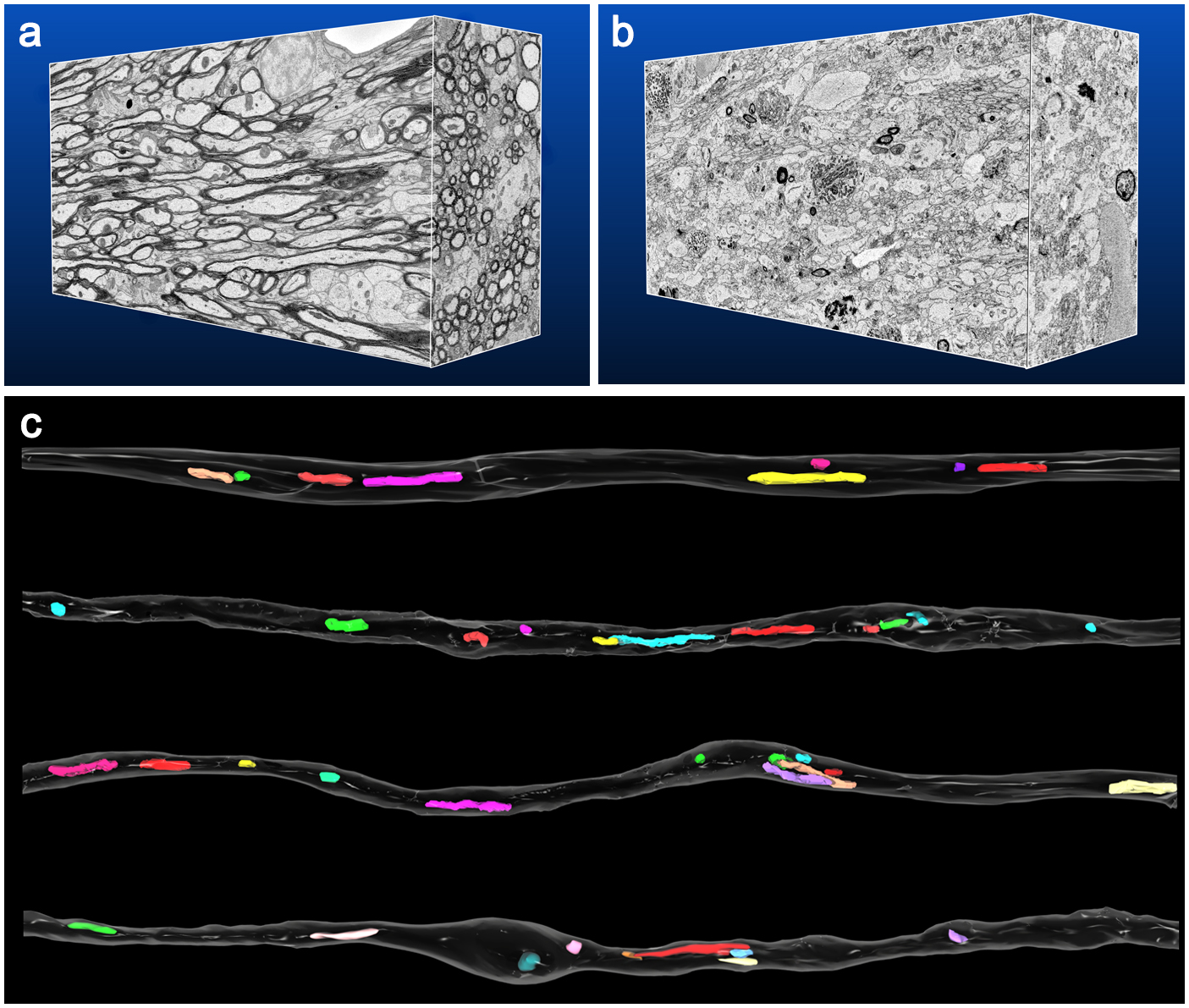
Figure 1. Reconstruction of serial electron microscopic images from corpus callosum of control (a) and demyelination model (b) mice, and 3D reconstrcution of axonal mitochondria (c). Modified from Ohno et al. PNAS (2014).
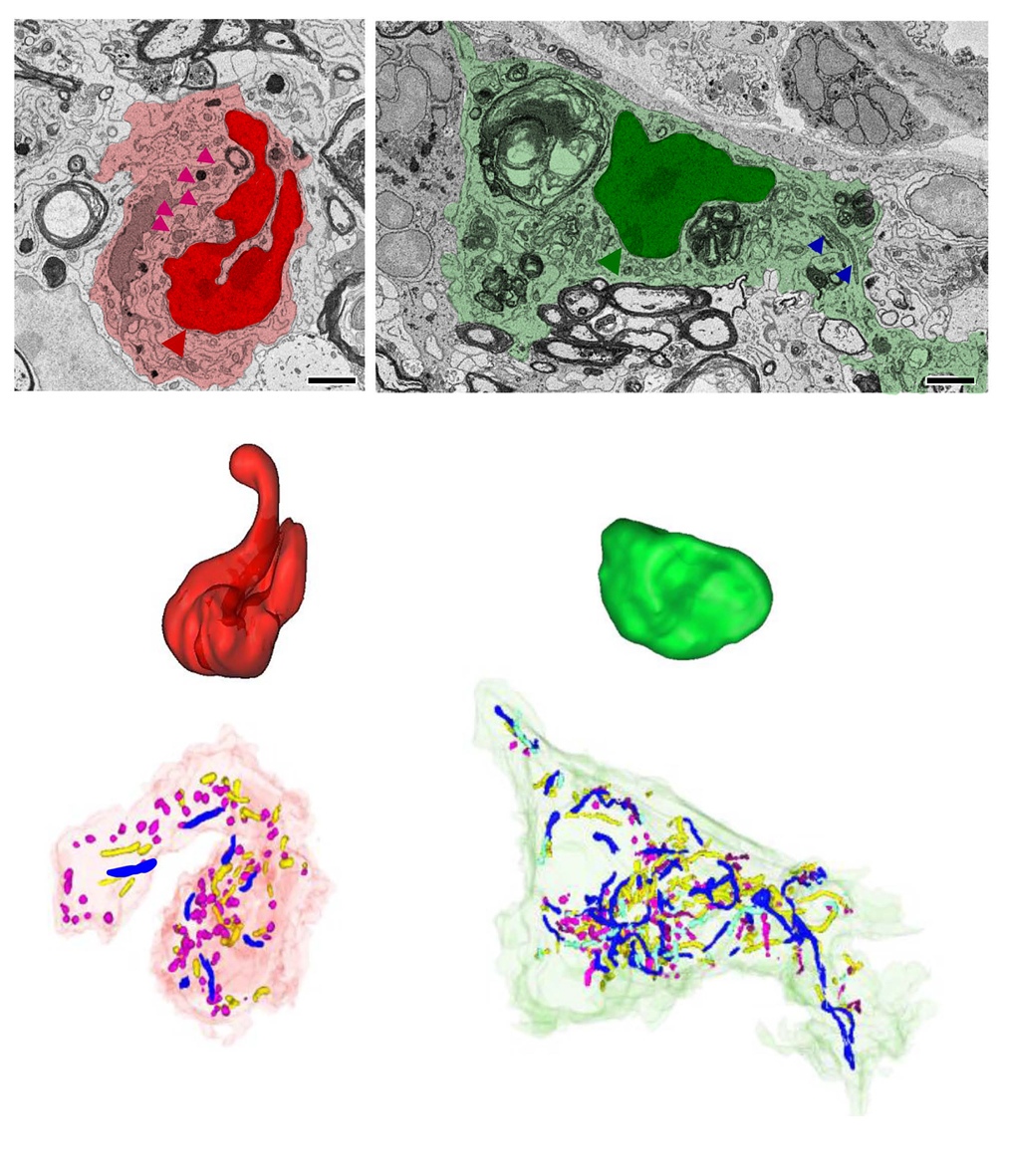
Figure 2. Colored electron microscopic images (upper row) and 3D reconstruction of nuclei (middle row) and mitochondria (lower row) of monocyte- (red) and microglia-derived (green) macrophages in a mouse spinal cord of a demyelination model. Modified from Katoh et al. Sci Rep (2017)


