We are leading the research and development of innovative bioimaging techniques and their application to basic medicine by utilizing state-of-the-art technologies such as laser optics and material chemistry. In particular, we are leading the world in the development of non-invasive imaging and manipulation techniques for live individuals and tissues, as well as super-resolution and ultra-high-speed imaging by utilizing multiphoton excitation and nonlinear optical processes. This has enabled us to develop quantitative visualization and analysis methods for physiological functions, and we aim to understand the principles of emergence and the molecular basis of life functions, including biological rhythms, through visualization and analysis of neural circuits and activities, and opening and release.
Recently, we have succeeded in developing a multiphoton microscope that enables fluorescence imaging of the deepest parts of living organisms, using near-infrared ultrashort pulse lasers and adaptive optics. In the brains of live mice, we succeeded in capturing tomographic images of nerve cells in the hippocampal dentate gyrus, which is approximately 1.6 mm deep from the surface, and also in observing the activity of hippocampal CA1 neurons at video rates. Furthermore, we are promoting research on biological rhythms in mammals, particularly the generation and function of circadian rhythms with a 24-hour cycle, using long-term imaging techniques for cellular functions.
On the other hand, we are also promoting the development of super-resolution microscopy techniques that can capture images of minute structures and molecular dynamics in living cells with a resolution approaching that of an electron microscope. Furthermore, using high-speed 3D imaging, we are applying it to the elucidation of the principles of emergence of local neural circuit functions, the physiological functions of endocrine and exocrine glands and plant cells, and the elucidation of the molecular mechanisms of disease onset.
In this research division, we are actively collaborating with a wide range of laboratories covering not only life sciences but also applied physics, material chemistry, basic medicine, and pharmacology, and are actively engaged in joint research. We aim to promote the creation of a new interdisciplinary field, with the advancement of imaging techniques that can capture physiological phenomena in living organisms as they are, and the cell physiology of nerves and secretion as the vertical and horizontal threads, respectively.
We are looking for graduate students and young researchers who are passionate about exploring new frontiers in science.
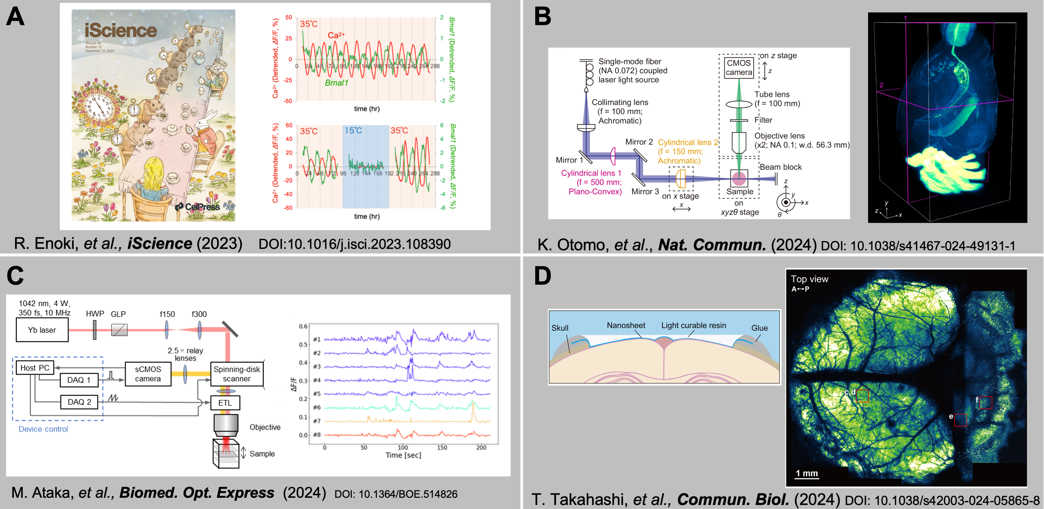
Fig.
(A) Cold-induced suspension and resetting of Ca2+ and transcriptional rhythms in the suprachiasmatic nucleus neurons. (B) Whole-brain fluorescence imaging of mouse using novel light-sheet microscopy. (C) High-speed volumetric imaging with an electronically tunable lens. (D) Ultra-widefield observation using novel optical materials.